You have no items in your shopping cart.
Featured
Description
Images & Validation
−Item 1 of 3
| Tested Applications | IF, IHC-P, WB |
|---|---|
| Dilution Range | IF - 1:25, WB - 1:2000, IHC-P - 1:10-50 |
| Reactivity | Human, Mouse |
Key Properties
−| Antibody Type | Primary Antibody |
|---|---|
| Host | Rabbit |
| Clonality | Polyclonal |
| Isotype | Rabbit IgG |
| Immunogen | Synthetic Peptide |
| Target | FOXD1 |
| Molecular Weight | 46140 |
| Conjugation | Unconjugated |
Storage & Handling
−| Storage | Maintain refrigerated at 2-8°C for up to 2 weeks. For long term storage store at -20°C in small aliquots to prevent freeze-thaw cycles |
|---|---|
| Form/Appearance | Purified polyclonal antibody supplied in PBS with 0.09% (W/V) sodium azide. This antibody is purified through a protein A column, followed by peptide affinity purification. |
| Expiration Date | 12 months from date of receipt. |
| Disclaimer | For research use only |
Alternative Names
−Anti-Forkhead box protein D1 antibody, anti-FOXD1 antibody, anti-FKHL8 antibody, anti-FREAC4 antibody
Quality Guarantee
Explore bioreagents carefree to elevate your research. All our products are rigorously tested for performance. If a product does not perform as described on its datasheet, our scientific support team will provide expert troubleshooting, a prompt replacement, or a refund. For full details, please see our Terms & Conditions and Buying Guide. Contact us at [email protected].
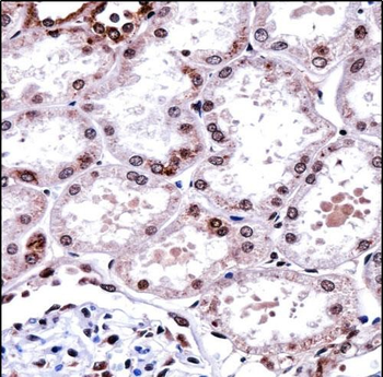
FOXD1 Antibody (N-term) immunohistochemistry analysis in formalin fixed and paraffin embedded human kidney tissue followed by peroxidase conjugation of the secondary antibody and DAB staining.This data demonstrates the use of FOXD1 Antibody (N-term) for immunohistochemistry. Clinical relevance has not been evaluated.
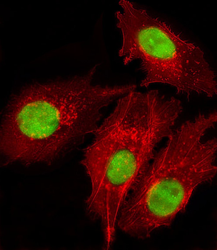
Immunofluorescent analysis of 4% paraformaldehyde-fixed, 0.1% Triton X-100 permeabilized HepG2 (human liver hepatocellular carcinoma cell line) cells labeling FOXD1 at 1/25 dilution, followed by Dylight 488-conjugated goat anti-rabbit IgG secondary antibody at 1/200 dilution (green). Immunofluorescence image showing nucleus staining on HepG2 cell line. Cytoplasmic actin is detected with Dylight 554 Phalloidin at 1/100 dilution (red).
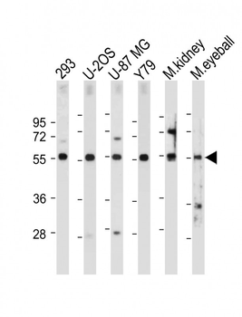
All lanes: Anti-FOXD1 Antibody (N-term) at 1:2000 dilution. Lane 1: 293 whole cell lysate. Lane 2: U-2OS whole cell lysate. Lane 3: U-87 MG whole cell lysate. Lane 4: Y79 whole cell lysate. Lane 5: Mouse kidney tissue lysate. Lane 6: Mouse eyeball tissue lysate. Lysates/proteins at 20 µg per lane. Secondary Goat Anti-Rabbit IgG, (H+L), Peroxidase conjugated at 1/10000 dilution. Predicted band size: 46 kDa. Blocking/Dilution buffer: 5% NFDM/TBST.
Quick Database Links
UniProt Details
− No UniProt data available
NCBI Reference Sequences
−Associated Accession Numbers
Curated reference sequences for the gene transcript and protein product| Protein | NP_004463.1 |
|---|
Documents Download
Datasheet
Product Information
Request a Document
Protocol Information
WB
Western Blot (IB, immunoblot)
IHC-P
Immunohistochemistry Paraffin
IF
Immunofluorescence
Filter by Applications
Filter by Species
Zhanwang Wang 1, Yi Jin 2, Dong He 3, Yuxing Zhu 1, Mengqing Xiao 1, Xiaoming Liu 4, Yaxin Cheng 1, Ke Cao Targeting ALG3/FOXD1/BNIP3 Axis Prevents Mitophagy and Gemcitabine Resistance of Nasopharyngeal Carcinoma Int J Biol Sci ., (2025)
Applications
IHC
Reactivity
Human
FOXD1 Antibody (N-term) (orb32533)

- 0.0
Based on 0 reviews
Participating in our Biorbyt product reviews program enables you to support fellow scientists by sharing your firsthand experience with our products.
Login to Submit a ReviewAvailable Sizes
Select a size below
Choose Conjugation or Carrier Free Version
Free Secondary Antibody (20 ul)0/0
Please add an antibody product to your cart first.