You have no items in your shopping cart.
Ferritin Heavy Chain Recombinant Rabbit Monoclonal Antibody
SKU: orb1607972
Featured
Description
Research Area
Alzheimer's Disease, Cardiovascuolar, Neurodegenerative Disease, Neuroscience, Vitamins and Minerals
Images & Validation
−Item 1 of 7
| Tested Applications | IF, IHC-Fr, IHC-P, WB |
|---|---|
| Dilution Range | WB=1:500-2000, IHC-P=1:100-500, IHC-F=1:100-500, IF=1:100-500 |
| Reactivity | Human |
Key Properties
−| Antibody Type | Primary Antibody |
|---|---|
| Host | Rabbit |
| Clonality | Recombinant |
| Isotype | IgG |
| Clone No. | B7D7 |
| Immunogen | A synthesized peptide derived from human Ferritin heavy chain (150-183aa) |
| Target | FTH1 |
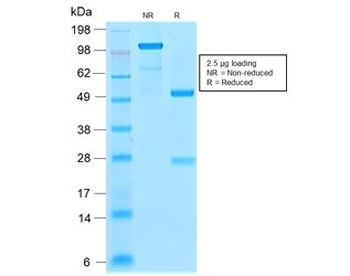
| Molecular Weight | 21 kDa |
| Purification | Affinity purified by Protein A |
| Conjugation | Unconjugated |
Customer Validated Data
− The data below is submitted by researchers worldwide. We provide this transparency to help you decide if this product works for your specific application or species, even if we haven't tested it ourselves.
Success by Application
Reactivity Distribution
Latest Experiments
4 ResultsDilution
Sample
Summary
WB
Mouse
1:1000
Hippocampal tissues from C57BL/6 mice
This antibody was used in Western...
WB
Mouse
-
Hippocampal tissues from db/db mice
This antibody was used in Western...
WB
Mouse
1:1000
Aortic homogenates from APOE⁻/⁻ mice
This antibody was used in Western...
WB, ELISA
Rat
-
Rat myocardial tissue (from SD rats) and H9C2 rat cardiomyoblast cells
This antibody was used in Western...
Storage & Handling
−| Storage | Maintain refrigerated at 2-8°C for up to 2 weeks. For long term storage store at -20°C in small aliquots to prevent freeze-thaw cycles. |
|---|---|
| Form/Appearance | Liquid |
| Buffer/Preservatives | 0.01M TBS (pH7.4) with 1% rAlbumin, 0.02% Proclin300 and 50% Glycerol. |
| Concentration | 1mg/ml |
| Expiration Date | 12 months from date of receipt. |
| Disclaimer | For research use only |
Alternative Names
−FTH, FTH1, FHC, FTHL6, HFE5, NBIA9, PIG15, PLIF, HFt, MFH, FRIH_FELCA, Ferritin H subunit, 1.16.3.1, FRIH_CANLF, FRIH_HUMAN, Cell proliferation-inducing gene 15 protein, FRIH_MOUSE, ferritin heavy chain 1, ferritin, heavy polypeptide 1, apoferritin, placenta immunoregulatory factor, proliferation-inducing protein 15, H-ferritin
Similar Products
−- Item 1 of 2
Recombinant Ferritin Light Chain Antibody / Rabbit Monoclonal [orb2640561]
IHC-P
Human
Rabbit
Recombinant
Unconjugated
7 ml
Quality Guarantee
Explore bioreagents carefree to elevate your research. All our products are rigorously tested for performance. If a product does not perform as described on its datasheet, our scientific support team will provide expert troubleshooting, a prompt replacement, or a refund. For full details, please see our Terms & Conditions and Buying Guide. Contact us at [email protected].

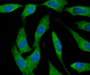
ICC staining of Ferritin Heavy Chain in Hela cells (green). Formalin fixed cells were permeabilized with 0.1% Triton X-100 in TBS for 10 minutes at room temperature and blocked with 1% Blocker BSA for 15 minutes at room temperature. Cells were probed with the primary antibody (orb1607972, 1/50) for 1 hour at room temperature, washed with PBS. Alexa Fluor®488 Goat anti-Rabbit IgG was used as the secondary antibody at 1/1000 dilution. The nuclear counter stain is DAPI (blue).
Immunohistochemical analysis of paraffin-embedded human brain tissue using anti-Ferritin Heavy Chain antibody. The section was pre-treated using heat mediated antigen retrieval with Tris-EDTA buffer (pH 8.0-8.4) for 20 minutes. The tissues were blocked in 5% BSA for 30 minutes at room temperature, washed with ddH2O and PBS, and then probed with the primary antibody (orb1607972, 1/50) for 30 minutes at room temperature. The detection was performed using an HRP conjugated compact polymer system. DAB was used as the chromogen. Tissues were counterstained with hematoxylin and mounted with DPX.
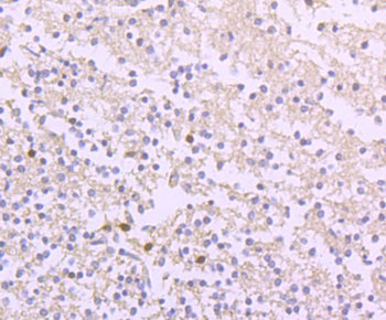
Immunohistochemical analysis of paraffin-embedded human brain tissue using anti-Ferritin Heavy Chain antibody. The section was pre-treated using heat mediated antigen retrieval with Tris-EDTA buffer (pH 8.0-8.4) for 20 minutes. The tissues were blocked in 5% BSA for 30 minutes at room temperature, washed with ddH2O and PBS, and then probed with the primary antibody (orb1607972, 1/50) for 30 minutes at room temperature. The detection was performed using an HRP conjugated compact polymer system. DAB was used as the chromogen. Tissues were counterstained with hematoxylin and mounted with DPX.06.
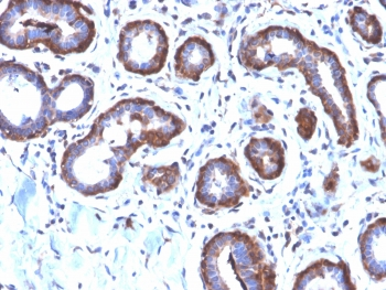
Immunohistochemical analysis of paraffin-embedded human liver tissue using anti-Ferritin Heavy Chain antibody. The section was pre-treated using heat mediated antigen retrieval with Tris-EDTA buffer (pH 8.0-8.4) for 20 minutes. The tissues were blocked in 5% BSA for 30 minutes at room temperature, washed with ddH2O and PBS, and then probed with the primary antibody (orb1607972, 1/50) for 30 minutes at room temperature. The detection was performed using an HRP conjugated compact polymer system. DAB was used as the chromogen. Tissues were counterstained with hematoxylin and mounted with DPX.
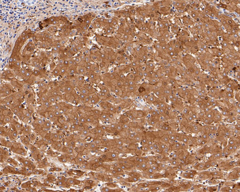
Immunohistochemical analysis of paraffin-embedded human liver tissue using anti-Ferritin Heavy Chain antibody. The section was pre-treated using heat mediated antigen retrieval with Tris-EDTA buffer (pH 8.0-8.4) for 20 minutes. The tissues were blocked in 5% BSA for 30 minutes at room temperature, washed with ddH2O and PBS, and then probed with the primary antibody (orb1607972, 1/50) for 30 minutes at room temperature. The detection was performed using an HRP conjugated compact polymer system. DAB was used as the chromogen. Tissues were counterstained with hematoxylin and mounted with DPX.

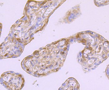
Immunohistochemical analysis of paraffin-embedded human placenta tissue using anti-Ferritin Heavy Chain antibody. The section was pre-treated using heat mediated antigen retrieval with Tris-EDTA buffer (pH 8.0-8.4) for 20 minutes. The tissues were blocked in 5% BSA for 30 minutes at room temperature, washed with ddH2O and PBS, and then probed with the primary antibody (orb1607972, 1/50) for 30 minutes at room temperature. The detection was performed using an HRP conjugated compact polymer system. DAB was used as the chromogen. Tissues were counterstained with hematoxylin and mounted with DPX.
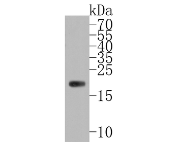
Western blot analysis of Ferritin Heavy Chain on human liver tissue lysates. Proteins were transferred to a PVDF membrane and blocked with 5% BSA in PBS for 1 hour at room temperature. The primary antibody (orb1607972, 1/500) was used in 5% BSA at room temperature for 2 hours. Goat Anti-Rabbit IgG - HRP Secondary Antibody at 1:5000 dilution was used for 1 hour at room temperature.
Quick Database Links
Gene Symbol
FTH1
UniProt
UniProt Details
− No UniProt data available
Documents Download
Datasheet
Product Information
Request a Document
Protocol Information
WB
Western Blot (IB, immunoblot)
IHC-P
Immunohistochemistry Paraffin
IHC-Fr
Immunohistochemistry Frozen
IF
Immunofluorescence
Ferritin Heavy Chain Recombinant Rabbit Monoclonal Antibody (orb1607972)

- 0.0
Based on 0 reviews
Participating in our Biorbyt product reviews program enables you to support fellow scientists by sharing your firsthand experience with our products.
Login to Submit a ReviewAvailable Sizes
Select a size below
Free Secondary Antibody (20 ul)0/0
Please add an antibody product to your cart first.