You have no items in your shopping cart.
Description
Research Area
Cancer Biology, Metabolism Research, Signal Transduction
Images & Validation
−Item 1 of 3
| Tested Applications | FC, IF, WB |
|---|---|
| Dilution Range | IF - 1:10-50, WB - 1:1000, FC - 1:10-50 |
| Reactivity | Human, Mouse |
| Predicted Reactivity | Bovine, Monkey |
Key Properties
−| Host | Rabbit |
|---|---|
| Clonality | Polyclonal |
| Isotype | Rabbit IgG |
| Immunogen | This ENOA antibody is generated from rabbits immunized with a KLH conjugated synthetic peptide between 33-60 amino acids from the N-terminal region of human ENOA. Antigen Region: 33-60 aa. |
| Target | ENO1 |
| Molecular Weight | 47169 Da |
| Conjugation | Unconjugated |
Storage & Handling
−| Storage | Maintain refrigerated at 2-8°C for up to 2 weeks. For long term storage store at -20°C in small aliquots to prevent freeze-thaw cycles |
|---|---|
| Form/Appearance | Purified polyclonal antibody supplied in PBS with 0.09% (W/V) sodium azide. This antibody is prepared by Saturated Ammonium Sulfate (SAS) precipitation followed by dialysis against PBS. |
| Expiration Date | 12 months from date of receipt. |
| Disclaimer | For research use only |
Alternative Names
−Alpha-enolase, 2-phospho-D-glycerate hydro-lyase, C-myc promoter-binding protein, Enolase 1, MBP-1, MPB-1, Non-neural enolase, NNE, Phosphopyruvate hydratase, Plasminogen-binding protein, ENO1, ENO1L1, MBPB1, MPB1
Similar Products
−- Item 1 of 3
ENOA Antibody (N-term) [orb1788344]
FC, IF, WB
Human, Mouse, Rat
Rabbit
Polyclonal
Unconjugated
Quality Guarantee
Explore bioreagents carefree to elevate your research. All our products are rigorously tested for performance. If a product does not perform as described on its datasheet, our scientific support team will provide expert troubleshooting, a prompt replacement, or a refund. For full details, please see our Terms & Conditions and Buying Guide. Contact us at [email protected].
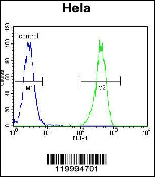
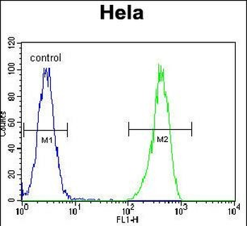
ENOA Antibody (N-term) flow cytometric analysis of Hela cells (right histogram) compared to a negative control cell (left histogram). FITC-conjugated goat-anti-rabbit secondary antibodies were used for the analysis.

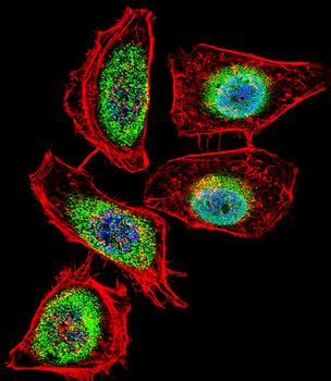
Fluorescent confocal image of Hela cell stained with ENOA Antibody (N-term).Hela cells were fixed with 4% PFA (20 min), permeabilized with Triton X-100 (0.1%, 10 min), then incubated with ENOA primary antibody (1:25, 1 h at 37°C). For secondary antibody, Alexa Fluor 488 conjugated donkey anti-rabbit antibody (green) was used (1:400, 50 min at 37°C).Cytoplasmic actin was counterstained with Alexa Fluor 555 (red) conjugated Phalloidin (7units/ml, 1 h at 37°C). Nuclei were counterstained with DAPI (blue) (10 µg/ml, 10 min). ENOA immunoreactivity is localized to Cytoplasm and Nucleus significantly.
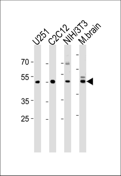
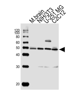
ENOA Antibody (N-term) western blot analysis in U251, mouse C2C12, mouse NIH/3T3 cell line and mouse brain tissue lysates (35 ug/lane). This demonstrates the ENOA antibody detected the ENOA protein (arrow).
Quick Database Links
UniProt Details
− No UniProt data available
NCBI Reference Sequences
−Associated Accession Numbers
Curated reference sequences for the gene transcript and protein product| Protein | NP_001188412.1, NP_001419.1 |
|---|
Documents Download
Datasheet
Product Information
Request a Document
Protocol Information
WB
Western Blot (IB, immunoblot)
FC
Flow Cytometry
IF
Immunofluorescence
ENOA Antibody (N-term) (orb1931371)

- 0.0
Based on 0 reviews
Participating in our Biorbyt product reviews program enables you to support fellow scientists by sharing your firsthand experience with our products.
Login to Submit a ReviewAvailable Sizes
Select a size below
Choose Conjugation or Carrier Free Version
Free Secondary Antibody (20 ul)0/0
Please add an antibody product to your cart first.