You have no items in your shopping cart.
Featured
KO/KD
Validated
Validated
Description
Research Area
Cancer Biology, Cell Biology
Images & Validation
−Item 1 of 14
| Tested Applications | ELISA, IF, IHC-P, KO/KD Validated, WB |
|---|---|
| Reactivity | Human, Mouse, Rat |
Key Properties
−| Antibody Type | Primary Antibody |
|---|---|
| Host | Rabbit |
| Clonality | Polyclonal |
| Isotype | IgG |
| Immunogen | Anti-EndoG antibody (orb1239381) was raised against a peptide corresponding to 15 amino acids near the amino terminus of human ENDOG. The immunogen is located within amino acids 40-90 of EndoG. |
| Target | ENDOG |
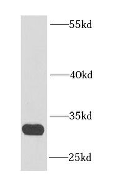
| Molecular Weight | Predicted: 33kDObserved: 33kD |
| Purification | EndoG Antibody is affinity chromatography purified via peptide column. |
| Conjugation | Unconjugated |
Storage & Handling
−| Storage | Maintain refrigerated at 2-8°C for up to 2 weeks. For long term storage store at -20°C in small aliquots to prevent freeze-thaw cycles. |
|---|---|
| Form/Appearance | Liquid |
| Buffer/Preservatives | EndoG Antibody is supplied in PBS containing 0.02% sodium azide. |
| Concentration | 1 mg/mL |
| Expiration Date | 12 months from date of receipt. |
| Disclaimer | For research use only |
Alternative Names
−EndoG Antibody: Endonuclease G, mitochondrial, Endo G
Similar Products
−- Item 1 of 5
Endo G Rabbit Polyclonal Antibody [orb6003]
IF, IHC-Fr, IHC-P, WB
Bovine, Canine, Equine, Guinea pig, Human, Porcine, Rabbit, Sheep
Mouse, Rat
Rabbit
Polyclonal
Unconjugated
50 μl, 100 μl, 200 μl - Item 1 of 1
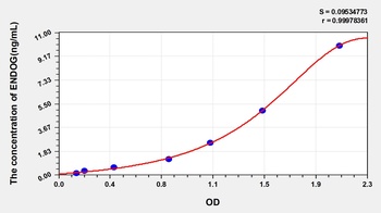
Human Endonuclease G, Mitochondrial (ENDOG) ELISA Kit [orb778073]
Human
0.16-10 ng/mL
0.065 ng/mL
48 T, 96 T - Item 1 of 1
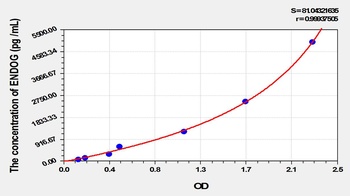
Rat Endonuclease G, Mitochondrial (ENDOG) ELISA Kit [orb781221]
Rat
78.13-5000 pg/mL
31 pg/mL
48 T, 96 T - Item 1 of 2
- Item 1 of 2
ENDOG Rabbit Polyclonal Antibody [orb626727]
ELISA, IHC, WB
Human, Mouse, Rat
Rabbit
Polyclonal
Unconjugated
50 μg, 100 μg


Quality Guarantee
Explore bioreagents carefree to elevate your research. All our products are rigorously tested for performance. If a product does not perform as described on its datasheet, our scientific support team will provide expert troubleshooting, a prompt replacement, or a refund. For full details, please see our Terms & Conditions and Buying Guide. Contact us at [email protected].
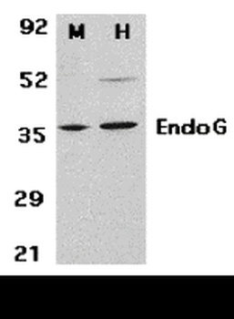
Western Blot Validation in Mouse 3T3 (M) and Human HepG2 (H) Cell Lysates. Loading: 15 µg of lysates per lane. Antibodies: EndoG orb1239381 (2 µg/mL), 1h incubation at RT in 5% NFDM/TBST. Secondary: Goat anti-rabbit IgG HRP conjugate at 1:10000 dilution.
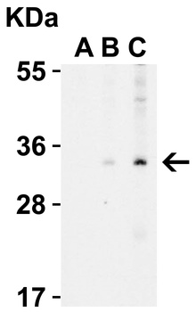
Western Blot Validation in Human A431 Cell Lysate with the presence (A) or absence (B and C) of blocking peptide. Loading: 15 µg of lysates per lane. Antibodies: EndoG orb1239381 (A: 0.5 µg/mL, B: 0.5 µg/mL, C: 1 µg/mL), 1h incubation at RT in 5% NFDM/TBST. Secondary: Goat anti-rabbit IgG HRP conjugate at 1:10000 dilution.
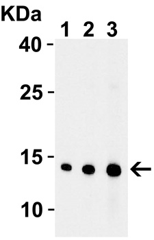
Western Blot Validation with Recombinant Protein. Loading: 30 ng of human EndoG recombinant protein per lane. Antibodies: EndoG orb1239381, 1h incubation at RT in 5% NFDM/TBST. Secondary: Goat anti-rabbit IgG HRP conjugate at 1:10000 dilution. Lane 1: 0.5 µg/mL, Lane 2: 1 µg/mL, Lane 3: 2 µg/mL.
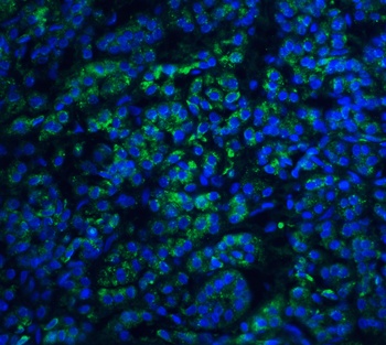
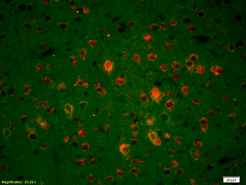
Immunofluorescence Validation of EndoG in Human Pancreas Tissue. Immunofluorescent analysis of 4% paraformaldehyde-fixed human pancreas tissue labeling EndoG with orb1239381 at 20 µg/mL, followed by goat anti-rabbit IgG secondary antibody at 1/500 dilution (green) and DAPI staining (blue).
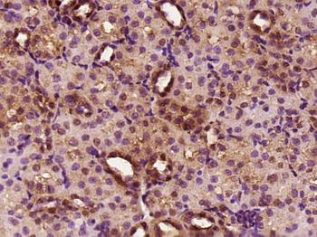
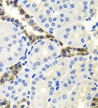
Immunohistochemistry Validation of EndoG in Human Pancreas Tissue. Immunohistochemical analysis of paraffin-embedded human pancreas tissue using anti-EndoG antibody (orb1239381) at 15 µg/ml. Tissue was fixed with formaldehyde and blocked with 10% serum for 1 h at RT; antigen retrieval was by heat mediation with a citrate buffer (pH6). Samples were incubated with primary antibody overnight at 4°C. A goat anti-rabbit IgG H&L (HRP) at 1/250 was used as secondary. Counter stained with Hematoxylin.
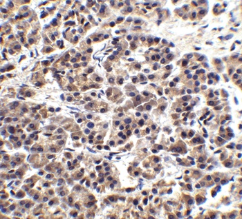
Immunohistochemistry Validation of EndoG in Human Pancreas Tissue. Immunohistochemical analysis of paraffin-embedded human pancreas tissue using anti-EndoG antibody (orb1239381) at 2.5 µg/ml. Tissue was fixed with formaldehyde and blocked with 10% serum for 1 h at RT; antigen retrieval was by heat mediation with a citrate buffer (pH6). Samples were incubated with primary antibody overnight at 4°C. A goat anti-rabbit IgG H&L (HRP) at 1/250 was used as secondary. Counter stained with Hematoxylin.
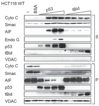
Localization Validation of EndoG by Purified p53 in Human Colorectal Cancer (HCT116) WT Cells (Wolff et al., 2008). Immunoblot analysis of subcellular fraction enriched with supernatant (S) was used to determine EndoG protein levels with increasing amounts of p53 (10, 20, 40, 100nM) in HCT116 WT cells. The release of EndoG from mitochondria induced by p53 is detected by anti-EndoG antibodies.
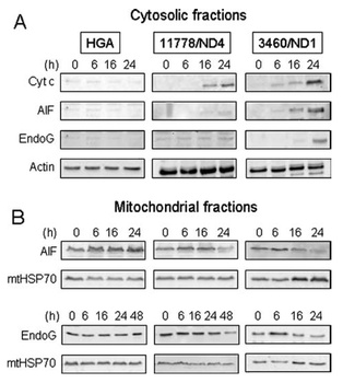
Localization Validation of EndoG in LHON cybrids (Zanna et al., 2005). Immunoblots of subcellular fractions enriched for (A) cytosol and (B) mitochondria were used to determine EndoG protein levels with anti-EndoG antibodies in LHON cybrids. The release of EndoG from mitochondria into the cytosol of 3460/ND1 mutant is observed after 24hr. Control (HGA) was unaffected.
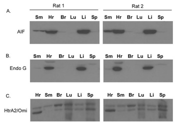
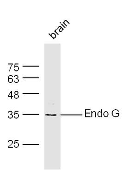
Tissue Specificity of EndoG in Rat Organs (Siu et al., 2007). WB analysis with anti-EndoG antibodies was performed for (B) EndoG in different organs of rats. EndoG was expressed very high in the heart and the liver of rats.
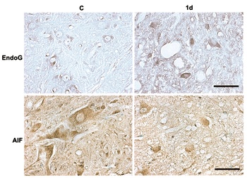
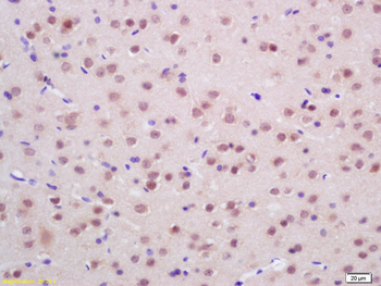
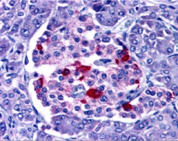
Immunohistochemistry Validation of EndoG translocation in Rat Spinal Cord Tissue (Yu et al., 2006). Protein analysis for EndoG translocation by immunohistochemistry with anti-EndoG antibodies in Rat spinal cord tissue. The staining showed that EndoG was expressed in the nuclei of SCI rats (1d) as compared to the perikarya (cytoplasm) in WT.
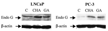
Translocation of EndoG by α-chaconine (CHA) and gallic acid (GA) in Human Prostate Cancer Cells (Reddivari et al., 2010). Lymph-node carcinoma of the prostate (LNCaP) or prostate cancer-3 (PC-3) cells were treated with CHA (2.5 µg/ml) or GA (15 µg/ml) for 24hr. EndoG expression detected by anti-EndoG antibodies was increased significantly in nuclear fraction after CHA or GA treatments while this effect was not observed in the solvent control (C).
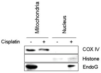
Translocation of EndoG by Cisplatin in Human HN4 cells (Kim et al., 2008). Head and Neck Squamous Cell Carcinoma (HNSCC), HN4 cells, from the patients were treated with 40 µM cisplatin. An increased expression of EndoG was induced in the nuclear fraction by cisplatin treatment, which was not observed in the mitochondria.
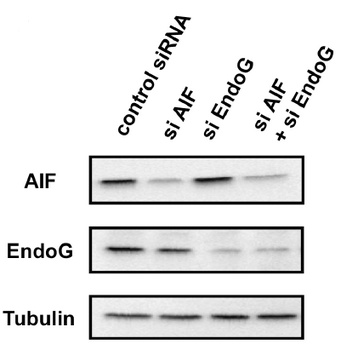
KD Validation of EndoG in Human small cell lung carcinoma Ms-1/Bcl-xL Cells (Sasazawa et al., 2009). Western blot analysis with anti-EndoG antibodies was performed for EndoG in Ms-1/Bcl-xL cells transfected with EndoG siRNA or control siRNA. EndoG expression was downregulated after EndoG siRNA knockdown.
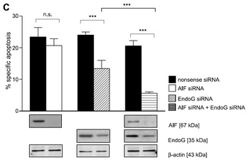
KD Validation of EndoG in Human MCF7 Cells (Schneiders et al., 2009). Western blot analysis with anti-EndoG antibodies was performed for EndoG in MCF7 cells transfected with EndoG siRNA or nonsense siRNA. EndoG expression was downregulated and cell apoptosis rate was remarkably decreased after EndoG siRNA knockdown.
Documents Download
Datasheet
Product Information
Request a Document
Protocol Information
WB
Western Blot (IB, immunoblot)
IHC-P
Immunohistochemistry Paraffin
IF
Immunofluorescence
ELISA
Enzyme-linked Immunosorbent Assay (EIA)
ENDOG Antibody (orb1239381)

- 0.0
Based on 0 reviews
Participating in our Biorbyt product reviews program enables you to support fellow scientists by sharing your firsthand experience with our products.
Login to Submit a ReviewAvailable Sizes
Select a size below
Free Secondary Antibody (20 ul)0/0
Please add an antibody product to your cart first.