You have no items in your shopping cart.
Description
Research Area
Epigenetics & Chromatin
Images & Validation
−Item 1 of 5
| Tested Applications | IHC-P, WB |
|---|---|
| Dilution Range | WB - 1:1000, IHC-P - 1:50-100 |
| Reactivity | Human, Mouse, Rat |
| Predicted Reactivity | Gallus |
Key Properties
−| Antibody Type | Primary Antibody |
|---|---|
| Host | Rabbit |
| Clonality | Polyclonal |
| Isotype | Rabbit IgG |
| Immunogen | This Dnmt3a antibody is generated from rabbits immunized with a KLH conjugated synthetic peptide between 457-486 amino acids from human Dnmt3a. Antigen Region: 457-486 aa. |
| Target | DNMT3A |
| Molecular Weight | 101858 Da |
| Conjugation | Unconjugated |
Storage & Handling
−| Storage | Maintain refrigerated at 2-8°C for up to 2 weeks. For long term storage store at -20°C in small aliquots to prevent freeze-thaw cycles |
|---|---|
| Form/Appearance | Purified polyclonal antibody supplied in PBS with 0.09% (W/V) sodium azide. This antibody is prepared by Saturated Ammonium Sulfate (SAS) precipitation followed by dialysis against PBS. |
| Expiration Date | 12 months from date of receipt. |
| Disclaimer | For research use only |
Alternative Names
−DNA (cytosine-5)-methyltransferase 3A, Dnmt3a, DNA methyltransferase HsaIIIA, DNA MTase HsaIIIA, MHsaIIIA, DNMT3A
Similar Products
−- Item 1 of 7
DNMT3A IP Mouse Monoclonal Antibody [orb2956662]
ELISA, FC, IF, IP
Human
Mouse
Monoclonal
Unconjugated
1 mg, 50 μg, 100 μg - Item 1 of 5
Dnmt3a Rabbit Polyclonal Antibody [orb10547]
FC, IF, IHC-Fr, IHC-P, WB
Bovine
Human, Mouse, Rat
Rabbit
Polyclonal
Unconjugated
50 μl, 100 μl, 200 μl - Item 1 of 1
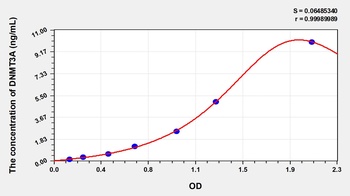
Human DNA Methyltransferase 3A (DNMT3A) ELISA Kit [orb778435]
Human
0.16-10 ng/mL
0.062 ng/mL
96 T, 48 T - Item 1 of 1
Mouse DNA Methyltransferase 3A (DNMT3A) ELISA Kit [orb1146948]
Mouse
0.16-10 ng/mL
0.062 ng/mL
48 T, 96 T - Item 1 of 5


Quality Guarantee
Explore bioreagents carefree to elevate your research. All our products are rigorously tested for performance. If a product does not perform as described on its datasheet, our scientific support team will provide expert troubleshooting, a prompt replacement, or a refund. For full details, please see our Terms & Conditions and Buying Guide. Contact us at [email protected].

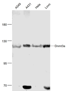
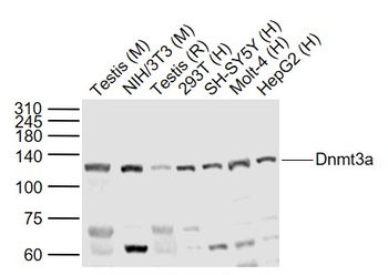
All lanes: Anti-Dnmt3a Antibody at 1:1000 dilution. Lane 1: Hela whole cell lysate. Lane 2: 293 whole cell lysate. Lane 3: Human skeletal muscle tissue lysate. Lane 4: NIH/3T3 whole cell lysate. Lane 5: Mouse brain tissue lysate. Lane 6: Rat heart tissue lysate. Lysates/proteins at 20 µg per lane. Secondary Goat Anti-Rabbit IgG, (H+L), Peroxidase conjugated at 1/10000 dilution. Predicted band size: 102 kDa. Blocking/Dilution buffer: 5% NFDM/TBST.
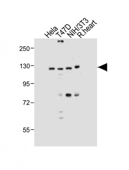
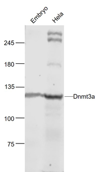
All lanes: Anti-Dnmt3a Antibody at 1:1000 dilution. Lane 1: Hela whole cell lysate. Lane 2: T47D whole cell lysate. Lane 3: NIH/3T3 whole cell lysate. Lane 4: Rat heart tissue lysate.Lysates/proteins at 20 µg per lane. Secondary Goat Anti-Rabbit IgG, (H+L), Peroxidase conjugated at 1/10000 dilution. Predicted band size: 102 kDa. Blocking/Dilution buffer: 5% NFDM/TBST.
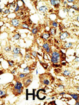
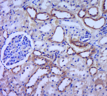
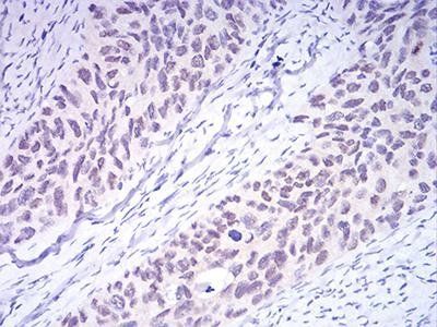
Formalin-fixed and paraffin-embedded human cancer tissue reacted with the primary antibody, which was peroxidase-conjugated to the secondary antibody, followed by DAB staining. This data demonstrates the use of this antibody for immunohistochemistry; clinical relevance has not been evaluated. BC = breast carcinoma; HC = hepatocarcinoma.
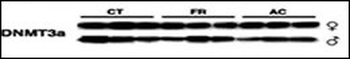
Lysates from mice thymus tissue after radiation were subjected to WB using antibody against DNMT3a. CT, control animals; FR, animals subjected to fractionated exposure; AC, acutely exposed animals. All sample loading was normalized to protein content. Representative Western blots from three independent experiments are shown; each lane represents a protein extract of a thymus of one animal. (Mol. Cancer Res. 2005 Oct 01;3 (10):553-561)
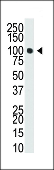
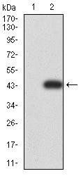
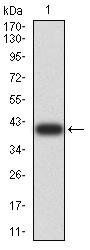
Western blot analysis of anti-Dnmt3a Pab in T47-D cell lysate. Dnmt3a (Arrow) was detected using purified Pab. Secondary HRP-anti-rabbit was used for signal visualization with chemiluminescence.
Quick Database Links
UniProt Details
− No UniProt data available
NCBI Reference Sequences
−Associated Accession Numbers
Curated reference sequences for the gene transcript and protein product| Protein | NP_783329.1, NP_072046.2, NP_715640.2, NP_783328.1 |
|---|
Documents Download
Datasheet
Product Information
Request a Document
Protocol Information
WB
Western Blot (IB, immunoblot)
IHC-P
Immunohistochemistry Paraffin
Dnmt3a Antibody (orb1939035)

- 0.0
Based on 0 reviews
Participating in our Biorbyt product reviews program enables you to support fellow scientists by sharing your firsthand experience with our products.
Login to Submit a ReviewAvailable Sizes
Select a size below
Choose Conjugation or Carrier Free Version
Free Secondary Antibody (20 ul)0/0
Please add an antibody product to your cart first.