You have no items in your shopping cart.
Featured
Description
Research Area
Signal Transduction
Images & Validation
−Item 1 of 7
| Tested Applications | ChIP, IF, IHC, IP, WB |
|---|---|
| Reactivity | Human, Mouse, Other, Rat |
| Application Notes |
Key Properties
−| Antibody Type | Primary Antibody |
|---|---|
| Host | Rabbit |
| Clonality | Polyclonal |
| Isotype | IgG |
| Immunogen | A synthetic peptide of human DiMethyl-Histone H3-K79 |
| Target | H3K79me2 |
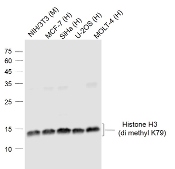
| Molecular Weight | Observed: 17kDa |
| Purification | Affinity purification |
| Conjugation | Unconjugated |
Storage & Handling
−| Storage | Maintain refrigerated at 2-8°C for up to 2 weeks. For long term storage store at -20°C in small aliquots to prevent freeze-thaw cycles. |
|---|---|
| Form/Appearance | Liquid |
| Buffer/Preservatives | PBS with 0.02% sodium azide, 50% glycerol, pH 7.3. |
| Concentration | batch dependent |
| Expiration Date | 12 months from date of receipt. |
| Disclaimer | For research use only |
Alternative Names
−H3F3A, H3t, H3.4, H3/g, H3FT
Similar Products
−- Item 1 of 13
Histone H3 (di methyl K79) Mouse Monoclonal Antibody [orb500704]
IF, IHC-Fr, IHC-P, WB
Mouse, Rat
Human, Mouse, Rat
Mouse
Monoclonal
Unconjugated
50 μl, 100 μl, 200 μl, 200 μg - Item 1 of 8
Histone H3 (di methyl K79) Mouse Monoclonal Antibody [orb499569]
IF, IHC-Fr, IHC-P, WB
Mouse, Rat
Human, Mouse, Rat
Mouse
Monoclonal
Unconjugated
50 μl, 100 μl, 200 μl, 200 μg 
Histone H3 (Di Methyl Lys79) rabbit pAb Antibody [orb2307801]
WB
Human, Mouse, Rat
Polyclonal
Unconjugated
50 μl, 100 μlHistone H3 (di methyl K79) Mouse Monoclonal Antibody (HRP) [orb474799]
IHC-Fr, IHC-P, WB
Human, Mouse, Rat
Mouse
Monoclonal
HRP
100 μlHistone H3 (di methyl K79) Mouse Monoclonal Antibody (Cy3) [orb976354]
IF
Human, Mouse, Rat
Mouse
Monoclonal
Cy3
100 μl
Quality Guarantee
Explore bioreagents carefree to elevate your research. All our products are rigorously tested for performance. If a product does not perform as described on its datasheet, our scientific support team will provide expert troubleshooting, a prompt replacement, or a refund. For full details, please see our Terms & Conditions and Buying Guide. Contact us at [email protected].
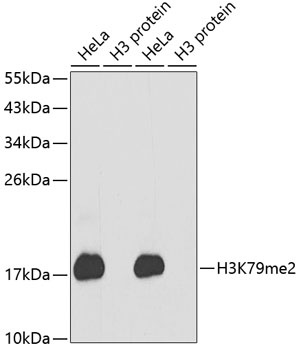
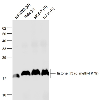
Western blot analysis of extracts of various cell lines, using DiMethyl-Histone H3-K79 antibody (orb1258180). Secondary antibody: HRP Goat Anti-Rabbit IgG (H+L) at 1:10000 dilution. Lysates/proteins: 25 ug per lane. Blocking buffer: 3% nonfat dry milk in TBST.
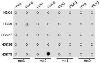
Dot-blot analysis of all sorts of methylation peptides using DiMethyl-Histone H3-K79 antibody (orb1258180).
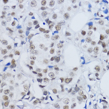
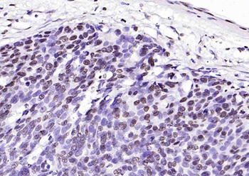
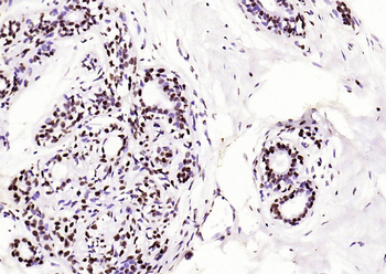
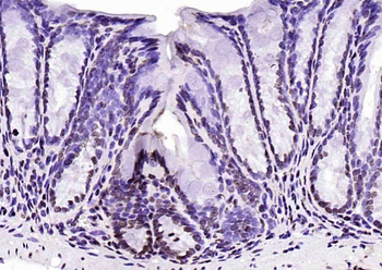
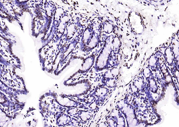
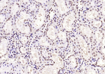
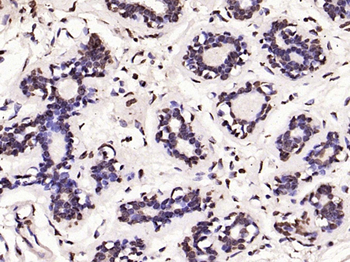
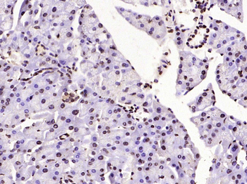
Immunohistochemistry of paraffin-embedded human mammary cancer using DiMethyl-Histone H3-K79 antibody (orb1258180) at dilution of 1:200 (40x lens).
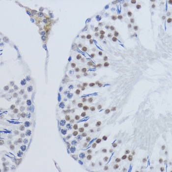
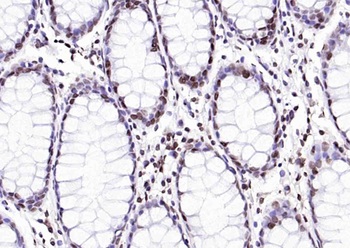
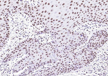
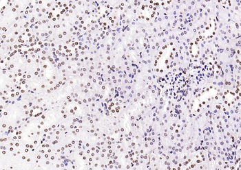
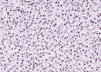
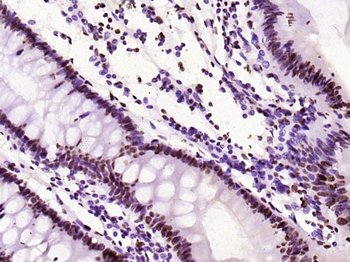
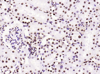
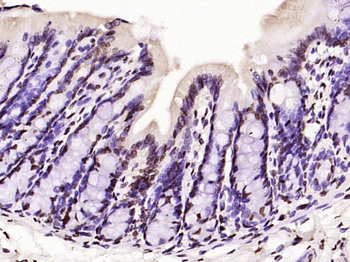
Immunohistochemistry of paraffin-embedded rat testis using DiMethyl-Histone H3-K79 antibody (orb1258180) at dilution of 1:200 (40x lens).
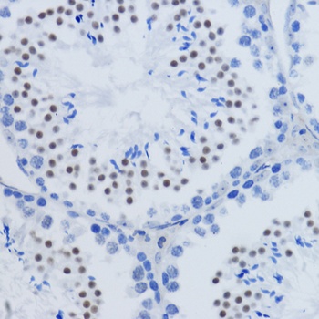
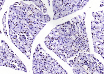
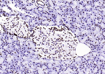
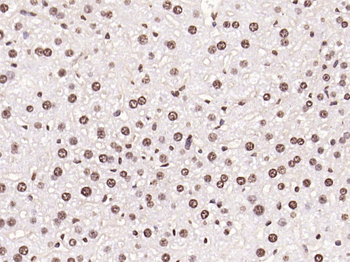
Immunohistochemistry of paraffin-embedded mouse testis using DiMethyl-Histone H3-K79 antibody (orb1258180) at dilution of 1:200 (40x lens).

Immunofluorescence analysis of 293T cells using DiMethyl-Histone H3-K79 antibody (orb1258180). Blue: DAPI for nuclear staining.
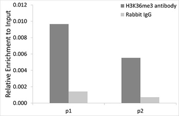
Chromatin Immunoprecipitation analysis of γ-actin gene from 293 cell line, using DiMethyl-Histone H3-K79 antibody (orb1258180) and rabbit IgG. P1, P2 and P3 were probes located on γ-actin gene as the schematic diagram illustrated. The amount of immunoprecipitated DNA was checked by quantitative PCR. Histogram was constructed by the ratios of the immunoprecipitated DNA to the input.
Quick Database Links
Gene Symbol
H3K79me2
UniProt
UniProt Details
− No UniProt data available
Documents Download
Datasheet
Product Information
Request a Document
Protocol Information
WB
Western Blot (IB, immunoblot)
IHC
Immunohistochemistry
IF
Immunofluorescence
IP
Immunoprecipitation
ChIP
Chromatin Immunoprecipitation
H3K79me2 Antibody (orb1258180)

- 0.0
Based on 0 reviews
Participating in our Biorbyt product reviews program enables you to support fellow scientists by sharing your firsthand experience with our products.
Login to Submit a ReviewAvailable Sizes
Select a size below
Choose Conjugation or Carrier Free Version
Free Secondary Antibody (20 ul)0/0
Please add an antibody product to your cart first.