You have no items in your shopping cart.
Description
Research Area
Musculoskeletal & Connective Tissue Research
Images & Validation
−Item 1 of 3
| Tested Applications | ELISA, IHC-P |
|---|---|
| Dilution Range | IHC-P: 1:100-500 (based on 0.5mg/ml) |
| Reactivity | Human, Mouse, Rat |
Key Properties
−| Host | Rabbit |
|---|---|
| Clonality | Polyclonal |
| Isotype | IgG |
| Immunogen | KLH conjugated synthetic peptide derived from human Collagen I. Please contact us for the exact immunogen sequence. The peptide is available as orb374938. |
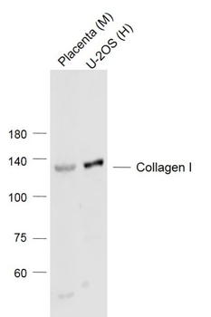
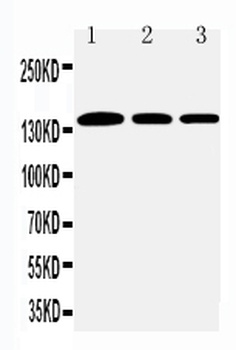
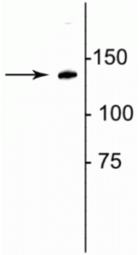
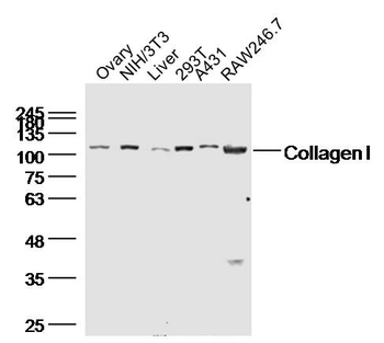
| Target | Collagen I |
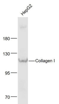
| Molecular Weight | 141kDa |
| Purity | Polyclonal antibodies are purified by peptide affinity chromatography |
| Conjugation | Unconjugated |
Storage & Handling
−| Storage | Maintain refrigerated at 2-8°C for up to 2 weeks. For long term storage store at -20°C in small aliquots to prevent freeze-thaw cycles. |
|---|---|
| Form/Appearance | 10 mM PBS, 0.02% sodium azide |
| Concentration | - 100 μg (in 200 μl): 0.5 mg/ml- 200 μg (in 400 μl): 0.5 mg/ml |
| Expiration Date | 12 months from date of receipt. |
| Disclaimer | For research use only |
Alternative Names
−Anti-CO1A1_HUMAN antibody, anti-Collagen I antibody, anti-COL1A1 antibody, anti-COL1A2 antibody, anti-Collagen type I alpha 1 antibody, anti-Collagen type I alpha 2 antibody
Similar Products
−- Item 1 of 8
Collagen I Rabbit Polyclonal Antibody [orb312178]
IF, IHC-Fr, IHC-P, WB
Bovine, Canine, Gallus, Sheep
Human, Mouse, Rabbit, Rat
Rabbit
Polyclonal
Unconjugated
- Item 1 of 11
Collagen I/COL1A1 Rabbit Polyclonal Antibody [orb371672]
IF, IHC, WB
Human
Rabbit
Polyclonal
Unconjugated
100 μg - Item 1 of 8
Collagen I/COL1A1 Rabbit Polyclonal Antibody [orb107158]
ICC, IF, IHC, WB
Human, Mouse, Rat
Rabbit
Polyclonal
Unconjugated
100 μg - Item 1 of 6
Collagen 1, alpha 1 telopeptide Antibody [orb319485]
ELISA, IHC, WB
Aves, Bovine, Canine, Equine, Feline, Gallus, Goat, Guinea pig, Hamster, Primate, Rabbit, Rodent, Xenopus
Human, Mouse, Rat, Sheep
Rabbit
Polyclonal
Unconjugated
100 μl - Item 1 of 4
Collagen I Rabbit Polyclonal Antibody [orb182761]
FC, ICC, IF, IHC-Fr, IHC-P, WB
Bovine, Canine, Gallus, Rabbit, Sheep
Human, Mouse, Rat
Rabbit
Polyclonal
Unconjugated
50 μl, 100 μl, 200 μl


Quality Guarantee
Explore bioreagents carefree to elevate your research. All our products are rigorously tested for performance. If a product does not perform as described on its datasheet, our scientific support team will provide expert troubleshooting, a prompt replacement, or a refund. For full details, please see our Terms & Conditions and Buying Guide. Contact us at [email protected].
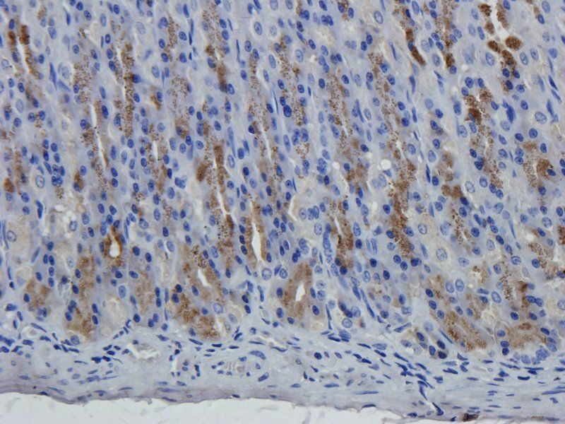
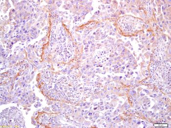
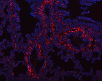
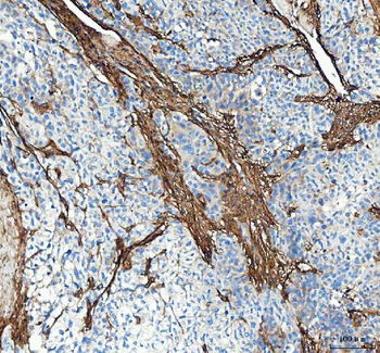
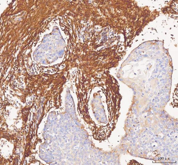
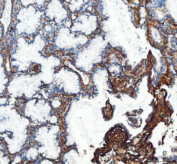
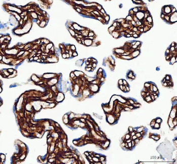
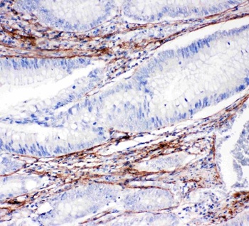
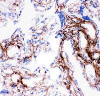
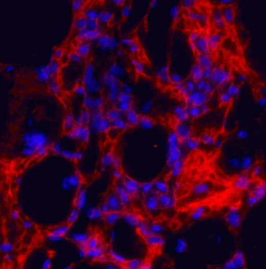
IHC-P staining of rat stomach tissue using Collagen I antibody (2.5 ug/ml)

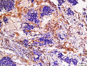
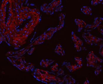
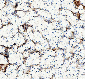
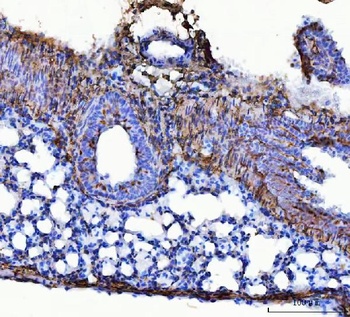
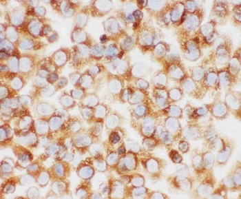
IHC-P image of mouse lymph node tissue using Collagen I antibody (5 ug/ml)
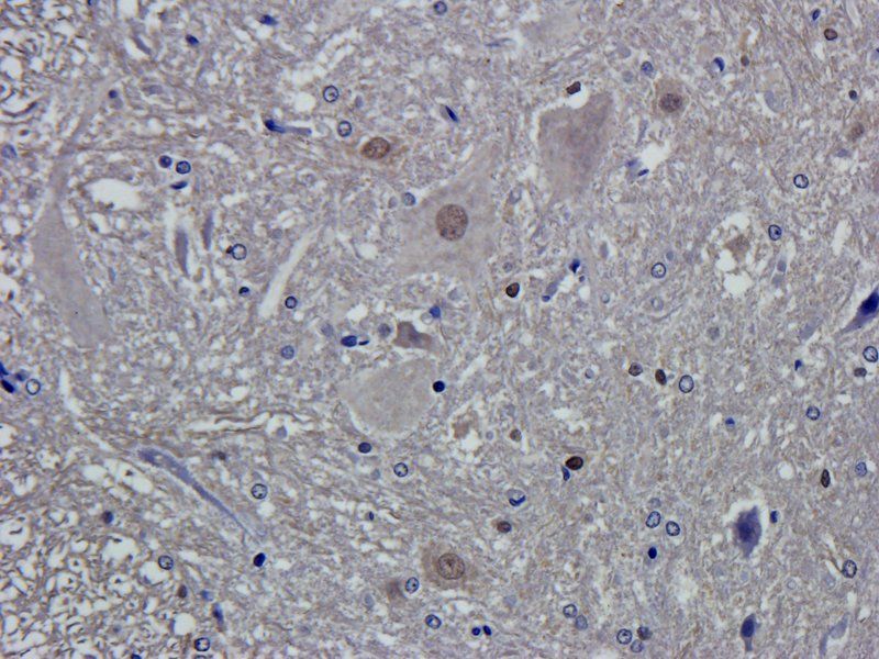
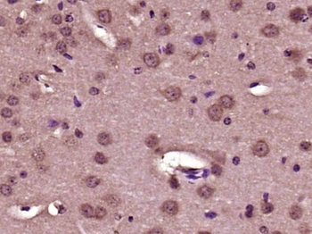
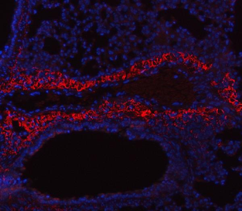
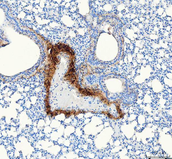
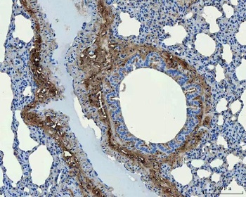
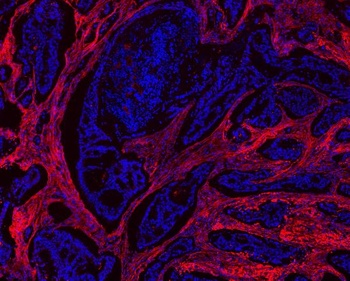
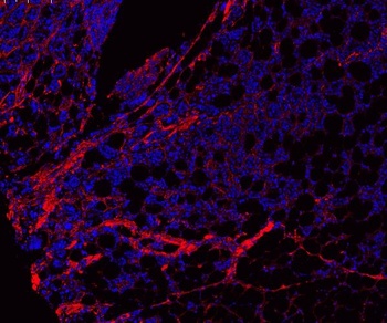
IHC-P staining of rat spinal cord tissue using anti-Collagen I (2.5 ug/ml)
Documents Download
Datasheet
Product Information
Request a Document
Protocol Information
IHC-P
Immunohistochemistry Paraffin
ELISA
Enzyme-linked Immunosorbent Assay (EIA)
Collagen I Rabbit Polyclonal Antibody (orb322979)

- 0.0
Based on 0 reviews
Participating in our Biorbyt product reviews program enables you to support fellow scientists by sharing your firsthand experience with our products.
Login to Submit a ReviewAvailable Sizes
Select a size below
Choose Conjugation or Carrier Free Version
Free Secondary Antibody (20 ul)0/0
Please add an antibody product to your cart first.