You have no items in your shopping cart.
Description
Research Area
Neuroscience
Images & Validation
−Item 1 of 5
| Tested Applications | FC, IF, IHC-P, WB |
|---|---|
| Dilution Range | IF - 1:25, WB - 1:500, IHC-P - 1:25, FC - 1:25 |
| Reactivity | Human, Mouse |
Key Properties
−| Host | Mouse |
|---|---|
| Clonality | Monoclonal |
| Isotype | IgG1,κ |
| Clone No. | B3646EV683X90X82 |
| Immunogen | This antibody is generated from a mouse immunized with a recombinant protein. Antigen Region: Recombinant Protein. |
| Target | CHRM2 |
| Molecular Weight | 51715 Da |
| Conjugation | Unconjugated |
Storage & Handling
−| Storage | Maintain refrigerated at 2-8°C for up to 2 weeks. For long term storage store at -20°C in small aliquots to prevent freeze-thaw cycles |
|---|---|
| Form/Appearance | Purified monoclonal antibody supplied in PBS with 0.09% (W/V) sodium azide. This antibody is purified through a protein G column, followed by dialysis against PBS. |
| Expiration Date | 12 months from date of receipt. |
| Disclaimer | For research use only |
Alternative Names
−Muscarinic acetylcholine receptor M2, CHRM2
Similar Products
−- Item 1 of 1
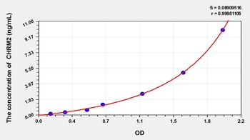
Mouse Cholinergic Receptor, Muscarinic 2 (CHRM2) ELISA Kit [orb777788]
Mouse
0.16-10 ng/mL
0.052 ng/mL
48 T, 96 T - Item 1 of 4
mAChR M2 rabbit pAb Antibody [orb767506]
ELISA, IF, WB
Human, Mouse, Rat
Polyclonal
Unconjugated
50 μl, 100 μl - Item 1 of 1
Human Cholinergic Receptor, Muscarinic 2 (CHRM2) ELISA Kit [orb775671]
Human
0.16-10 ng/mL
0.056 ng/mL
48 T, 96 T - Item 1 of 1
Rat Cholinergic Receptor, Muscarinic 2 (CHRM2) ELISA Kit [orb780439]
Rat
0.16-10 ng/mL
0.053 ng/mL
48 T, 96 T - Item 1 of 4
ChRM2 Rabbit Polyclonal Antibody [orb10388]
IF, IHC-Fr, IHC-P, WB
Gallus, Porcine
Human, Mouse, Rat
Rabbit
Polyclonal
Unconjugated
50 μl, 100 μl, 200 μl


Quality Guarantee
Explore bioreagents carefree to elevate your research. All our products are rigorously tested for performance. If a product does not perform as described on its datasheet, our scientific support team will provide expert troubleshooting, a prompt replacement, or a refund. For full details, please see our Terms & Conditions and Buying Guide. Contact us at [email protected].
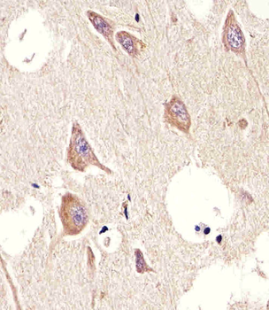
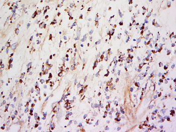
Immunohistochemical analysis of paraffin-embedded H. brain section using CHRM2. Diluted at 1:25 dilution. A undiluted biotinylated goat polyvalent antibody was used as the secondary, followed by DAB staining.
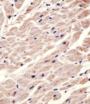
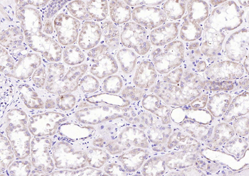
Immunohistochemical analysis of paraffin-embedded H. heart section using CHRM2. Diluted at 1:25 dilution. A undiluted biotinylated goat polyvalent antibody was used as the secondary, followed by DAB staining.
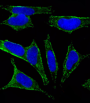
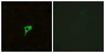
Fluorescent image of SH-SY5Y cells stained with CHRM2 Antibody. Diluted at 1:25 dilution. An Alexa Fluor 488-conjugated goat anti-mouse lgG at 1:400 dilution was used as the secondary antibody (green). DAPI was used to stain the cell nuclear (blue).
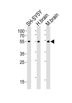
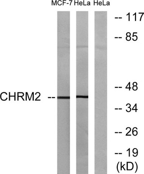
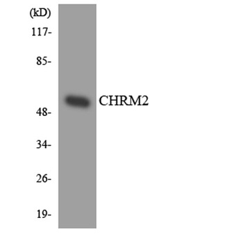
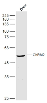
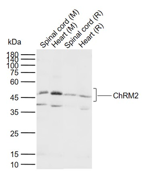
Western blot analysis of lysates from SH-SY5Y cell line, human brain, mouse brain tissue (from left to right), using CHRM2 Antibody. Diluted at 1:500 at each lane. A goat anti-mouse IgG H&L (HRP) at 1: 3000 dilution was used as the secondary antibody. Lysates at 20 μg per lane.
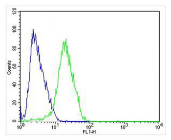
Overlay histogram showing SH-SY5Y cells stained (green line). The cells were fixed with 4% paraformaldehyde (10 min) and then permeabilized with 90% methanol for 10 min. The cells were then icubated in 2% bovine serum albumin to block non-specific protein-protein interactions followed by the antibody (, 1:25 dilution) for 60 min at 37°C. The secondary antibody used was Alexa Fluor 488 goat anti-mouse lgG (166821) at 1/200 dilution for 40 min at 37°C. Isotype control antibody (blue line) was mouse IgG1 (1 μg/1x10^6 cells) used under the same conditions. Acquisition of > 10000 events was performed.
Quick Database Links
Gene Symbol
CHRM2
UniProt
UniProt Details
− No UniProt data available
Documents Download
Datasheet
Product Information
Request a Document
Protocol Information
WB
Western Blot (IB, immunoblot)
IHC-P
Immunohistochemistry Paraffin
FC
Flow Cytometry
IF
Immunofluorescence
CHRM2 Antibody (orb1926763)

- 0.0
Based on 0 reviews
Participating in our Biorbyt product reviews program enables you to support fellow scientists by sharing your firsthand experience with our products.
Login to Submit a ReviewAvailable Sizes
Select a size below
Choose Conjugation or Carrier Free Version
Free Secondary Antibody (20 ul)0/0
Please add an antibody product to your cart first.