You have no items in your shopping cart.
Featured
Description
Research Area
Immunology & Inflammation; Cell Biology
Images & Validation
−Item 1 of 4
| Tested Applications | FACS, FC, ICC, IF, IHC, IP, WB |
|---|---|
| Dilution Range | WB (1:1000), IHC (1:100), ICC/IF (1:50); optimal dilutions for assays should be determined by the user. |
| Reactivity | Human, Mouse |
| Application Notes |
Key Properties
−| Host | Mouse |
|---|---|
| Clonality | Monoclonal |
| Isotype | IgG1 |
| Clone No. | PIN.1 |
| Immunogen | Human CD74 invariant chain synthetic peptide |
| Target | CD74 |
| Molecular Weight | 33-35kDa |
| Purification | Protein G Purified |
| Conjugation | Unconjugated |
Storage & Handling
−| Storage | Maintain refrigerated at 2-8°C for up to 2 weeks. For long term storage store at -20°C in small aliquots to prevent freeze-thaw cycles. |
|---|---|
| Buffer/Preservatives | PBS pH 7.2, 50% glycerol, 0.09% sodium azide. Storage buffer changes when conjugated. |
| Concentration | 1 mg/ml |
| Expiration Date | 12 months from date of receipt. |
| Disclaimer | For research use only |
Alternative Names
−CD74, DHLAG, HLA DR gamma, HLADG, p33, p35, Protein 41, CD74 antigen (invariant polypeptide of major histocompatibility complex, class II antigen-associated), CD74 antigen, CD74 molecule, CD74 molecule, major histocompatibility complex, class II invariant chain, CLIP, Gamma chain of class II antigens, HG2A_HUMAN, HLA class II histocompatibility antigen gamma chain, HLA DR antigens associated invariant chain, HLA-DR antigens-associated invariant chain, HLA-DR-gamma, HLADR antigens associated invariant chain, Ia antigen associated invariant chain, Ia antigen-associated invariant chain, Ia associated invariant chain, Ia gamma, Ii, Invariant polypeptide of major histocompatibility complex class II antigen associated, la-gamma, Major histocompatibility complex class II invariant chain, MHC HLA DR gamma chain, MHC HLA-DR gamma chain
Similar Products
−- Item 1 of 6
CD74 Rabbit Polyclonal Antibody [orb669066]
FC, ICC, IF, IHC
Human, Mouse, Rat
Rabbit
Polyclonal
Unconjugated
100 μg - Item 1 of 4
CD74 rabbit pAb Antibody [orb767069]
ELISA, IF, IHC, WB
Human, Mouse, Rat
Polyclonal
Unconjugated
100 μl - Item 1 of 5
- Item 1 of 5
- Item 1 of 5
CD74 Rabbit Polyclonal Antibody [orb1402197]
ELISA, FC, IF, IHC, WB
Human
Rabbit
Polyclonal
Unconjugated
100 μg


Quality Guarantee
Explore bioreagents carefree to elevate your research. All our products are rigorously tested for performance. If a product does not perform as described on its datasheet, our scientific support team will provide expert troubleshooting, a prompt replacement, or a refund. For full details, please see our Terms & Conditions and Buying Guide. Contact us at [email protected].
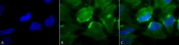
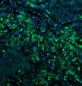
Immunocytochemistry/Immunofluorescence analysis using Mouse Anti-CD74 Monoclonal Antibody, Clone PIN 1.1. Tissue: Cervical cancer cell line (HeLa). Species: Human. Fixation: 2% Formaldehyde for 20 min at RT. Primary Antibody: Mouse Anti-CD74 Monoclonal Antibody at 1:100 for 12 hours at 4°C. Secondary Antibody: FITC Goat Anti-Mouse (green) at 1:200 for 2 hours at RT. Counterstain: DAPI (blue) nuclear stain at 1:40000 for 2 hours at RT. Localization: Cell membrane. Endoplasmic reticulum membrane. Golgi apparatus. Endosome. Lysosome. Magnification: 100x. (A) DAPI (blue) nuclear stain. (B) Anti-CD74 Antibody. (C) Composite.

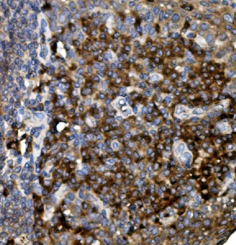
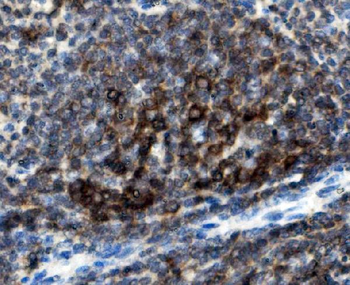
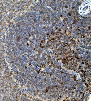
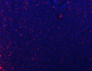
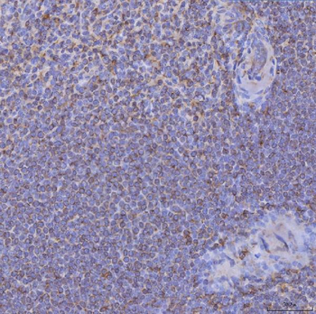
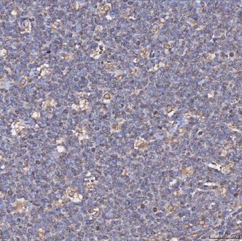
Immunohistochemistry analysis using Mouse Anti-CD74 Monoclonal Antibody, Clone PIN 1.1. Tissue: backskin. Species: Mouse. Fixation: Bouin's Fixative and paraffin-embedded. Primary Antibody: Mouse Anti-CD74 Monoclonal Antibody at 1:100 for 1 hour at RT. Secondary Antibody: FITC Goat Anti-Mouse (green) at 1:50 for 1 hour at RT. Localization: Beautiful basal to suprabasal staining in epidermis, dermis, hair follicles and muscle.
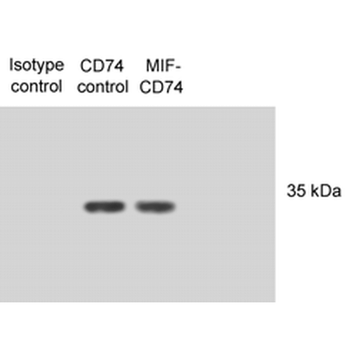
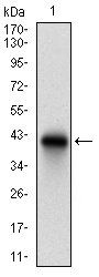
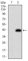
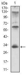
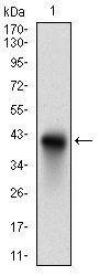
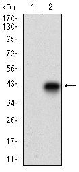
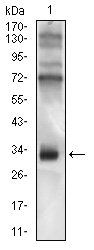
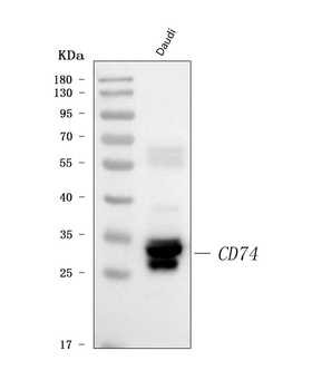
Western Blot analysis of Human N87 cell lysates showing detection of CD74 protein using Mouse Anti-CD74 Monoclonal Antibody, Clone PIN 1.1. Primary Antibody: Mouse Anti-CD74 Monoclonal Antibody at 1:1000. Lysates treated with macrophage inhibitory factor (MIF).
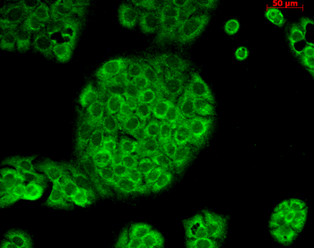
Immunocytochemistry/Immunofluorescence analysis using Mouse Anti-CD74 Monoclonal Antibody, Clone PIN 1.1. Tissue: HaCaT cells. Species: Human. Fixation: Cold 100% methanol for 10 minutes at -20°C. Primary Antibody: Mouse Anti-CD74 Monoclonal Antibody at 1:100 for 1 hour at RT. Secondary Antibody: FITC Goat Anti-Mouse (green) at 1:50 for 1 hour at RT. Localization: Cytoplasmic Staining.
Quick Database Links
UniProt Details
− No UniProt data available
NCBI Gene Details
− No NCBI Gene data available
NCBI Reference Sequences
−Associated Accession Numbers
Curated reference sequences for the gene transcript and protein product| Protein | NP_001020329.1 |
|---|
Documents Download
Datasheet
Product Information
Request a Document
Protocol Information
WB
Western Blot (IB, immunoblot)
IHC
Immunohistochemistry
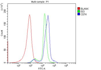
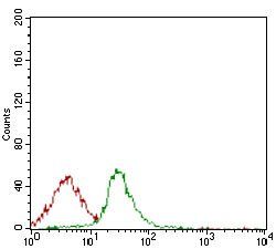
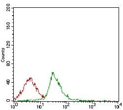
FACS
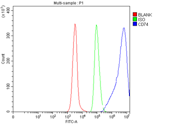
Fluorescence-Activated Cell Sorting (FC, Flow cytometry)
FC
Flow Cytometry
IF
Immunofluorescence
ICC
Immunocytochemistry
IP
Immunoprecipitation
CD74 Antibody (orb1822499)

- 0.0
Based on 0 reviews
Participating in our Biorbyt product reviews program enables you to support fellow scientists by sharing your firsthand experience with our products.
Login to Submit a ReviewAvailable Sizes
Select a size below
Choose Conjugation or Carrier Free Version
Free Secondary Antibody (20 ul)0/0
Please add an antibody product to your cart first.