You have no items in your shopping cart.
Featured
Description
Research Area
Immunology & Inflammation
Images & Validation
−Item 1 of 7
| Tested Applications | ELISA, ICC, IF, IHC-P, WB |
|---|---|
| Dilution Range | WB: 2 μg/ml, IHC-P:1:100, IF/ICC: 1:100 |
| Application Notes |
Key Properties
−| Host | Rabbit |
|---|---|
| Clonality | Polyclonal |
| Isotype | IgG |
| Immunogen | CD68 recombinant protein |
| Target | CD68 |
| Molecular Weight | 37 kDa |
| Purity | Polyclonal antibodies are purified by peptide affinity chromatography |
| Conjugation | Unconjugated |
Storage & Handling
−| Storage | Maintain refrigerated at 2-8°C for up to 2 weeks. For long term storage store at -20°C in small aliquots to prevent freeze-thaw cycles. |
|---|---|
| Form/Appearance | 10 mM PBS, 0.02% sodium azide |
| Concentration | - 100 μg (in 200 μl): 0.5 mg/ml- 200 μg (in 400 μl): 0.5 mg/ml |
| Expiration Date | 12 months from date of receipt. |
| Disclaimer | For research use only |
Alternative Names
−anti CD68 antibody, anti CD68gen antibody, anti CD68 molecule antibody, anti DKFZp686M18236 antibody, anti GP110 antibody, anti Macrophagegen CD68 (microsialin) antibody, anti macrosialin antibody, anti SCARD1 antibody, anti Scavenger receptor class D member 1 antibody
Similar Products
−- Item 1 of 11
CD68 Rabbit Polyclonal Antibody [orb10343]
IHC-P, WB
Human, Mouse, Rat
Rabbit
Polyclonal
Unconjugated
100 μg - Item 1 of 7
CD68 Rabbit Polyclonal Antibody [orb317599]
IF, IHC-Fr, IHC-P, WB
Rat
Mouse, Rat
Rabbit
Polyclonal
Unconjugated
50 μl, 100 μl, 200 μl - Item 1 of 7
CD68 Rabbit Polyclonal Antibody [orb500796]
IHC-Fr, IHC-P
Human
Human
Rabbit
Polyclonal
Unconjugated
50 μl, 100 μl - Item 1 of 8
- Item 1 of 4
CD68 Rabbit Polyclonal Antibody [orb10344]
WB
Bovine
Human, Mouse, Rat
Rabbit
Polyclonal
Unconjugated
50 μl, 100 μl, 200 μl


Quality Guarantee
Explore bioreagents carefree to elevate your research. All our products are rigorously tested for performance. If a product does not perform as described on its datasheet, our scientific support team will provide expert troubleshooting, a prompt replacement, or a refund. For full details, please see our Terms & Conditions and Buying Guide. Contact us at [email protected].
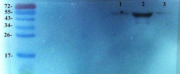
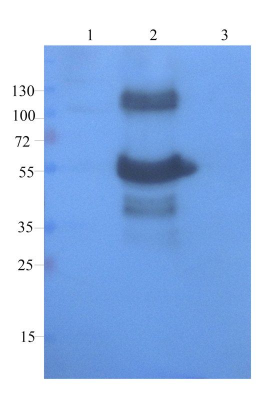
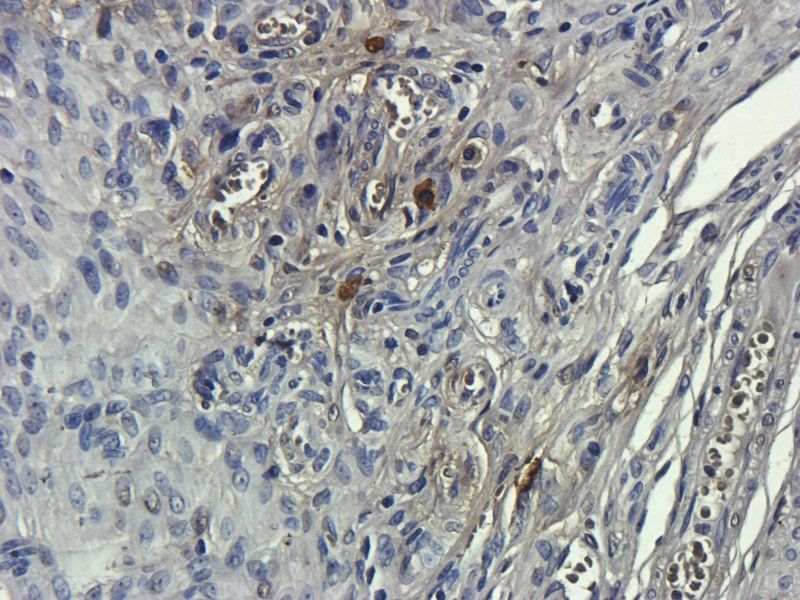
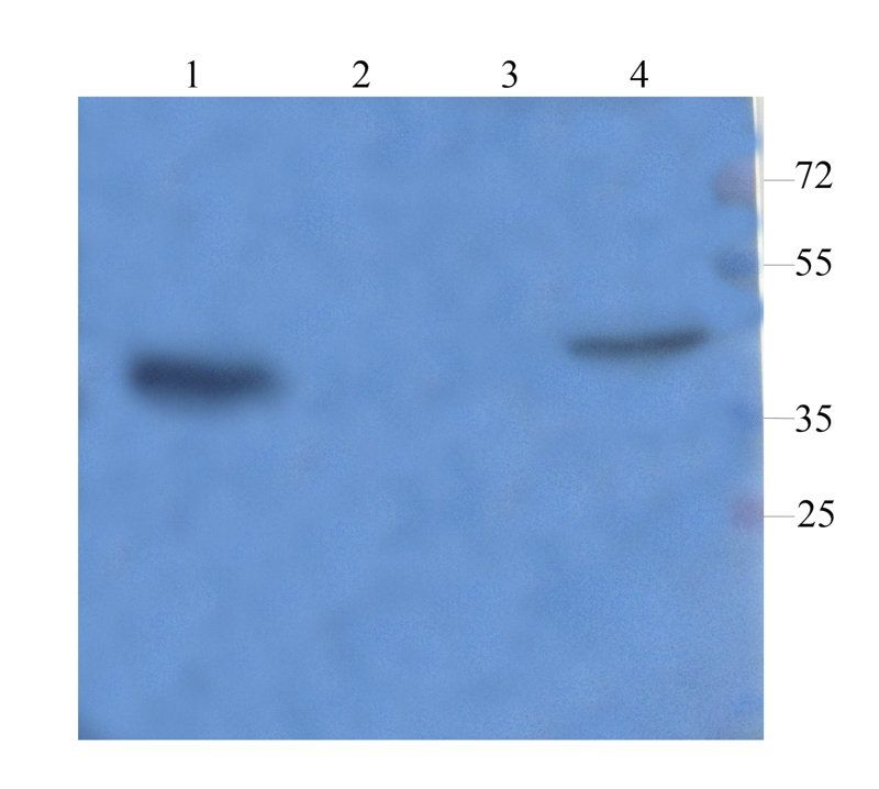
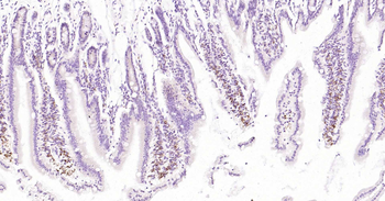
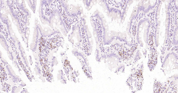
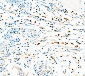
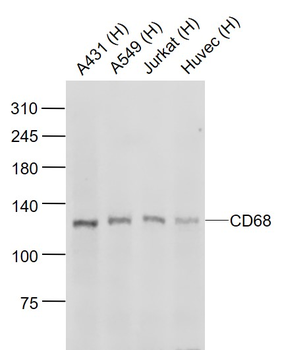
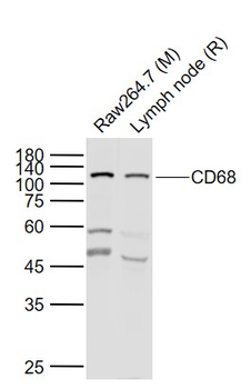
Western blot analysis of breast cancer (Lane 1), breast cancer (Lane 2), breast cancer (Lane 3) using CD68 antibody (dilution at 2 ug/ml)
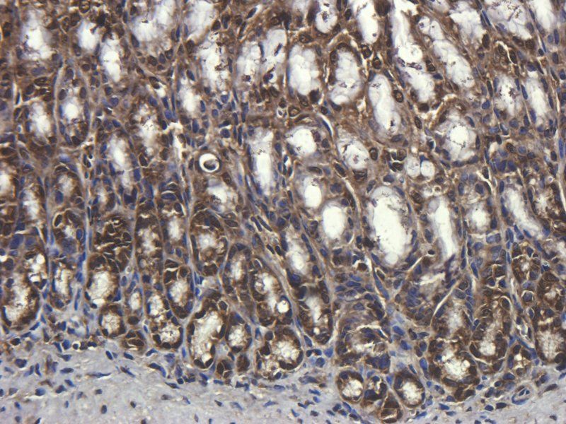
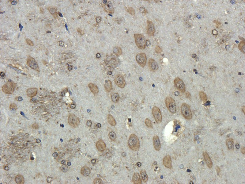
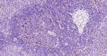
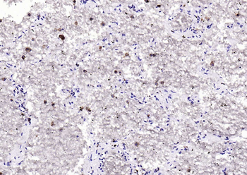
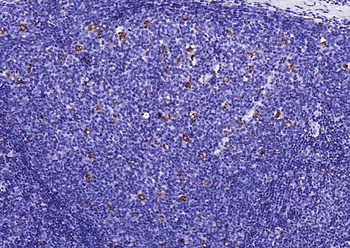
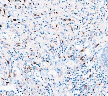
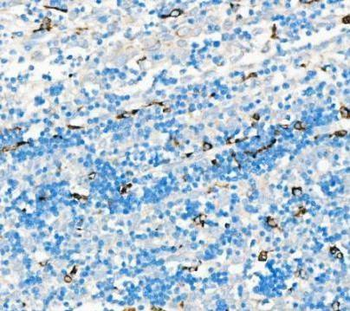
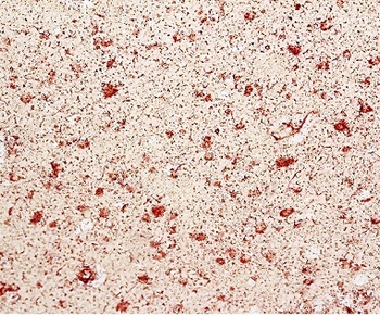
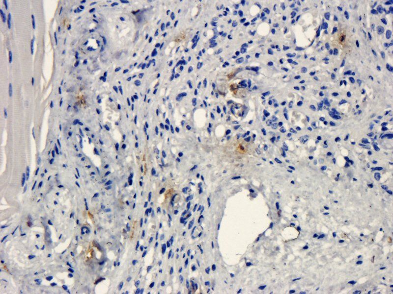
Immunohistochemical staining of rat spleen tissue using CD68 antibody (dilution of primary antibody - 1:100)
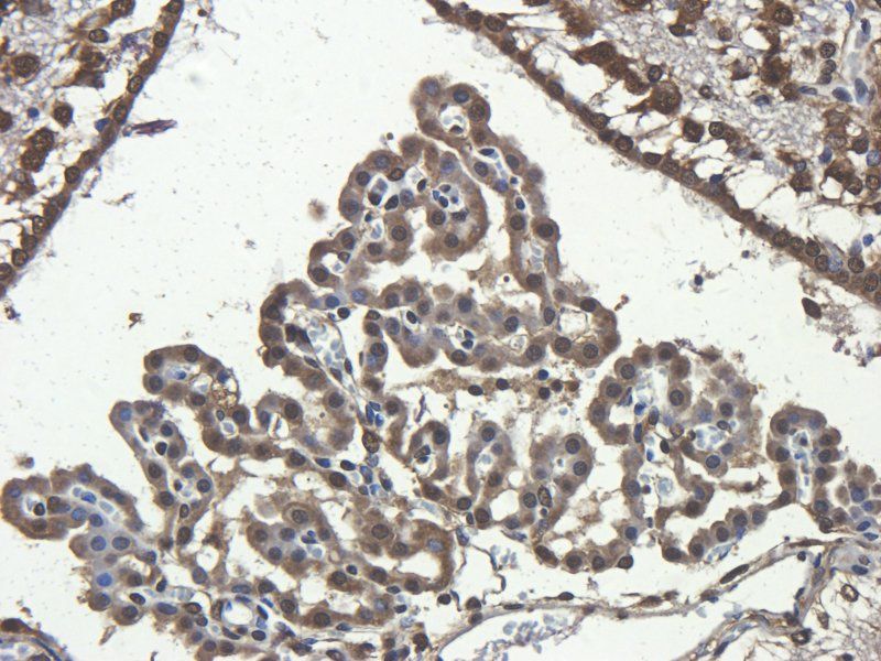
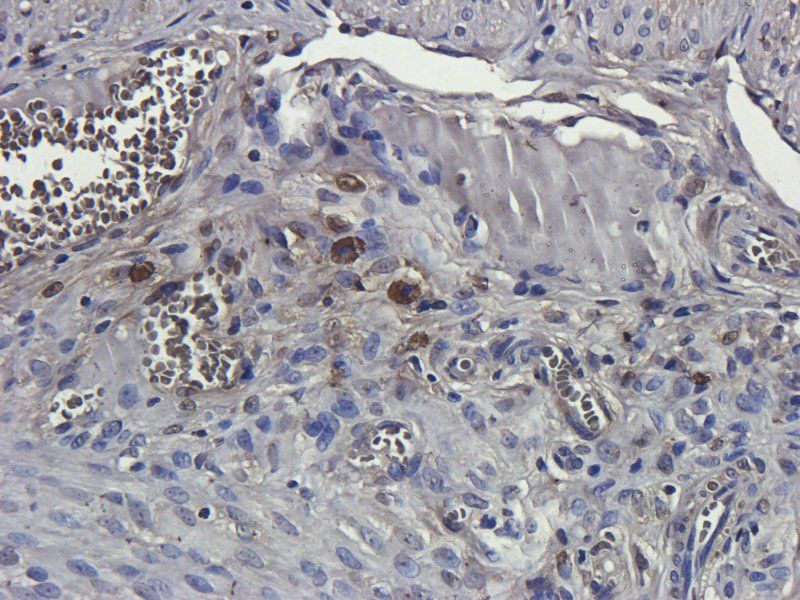
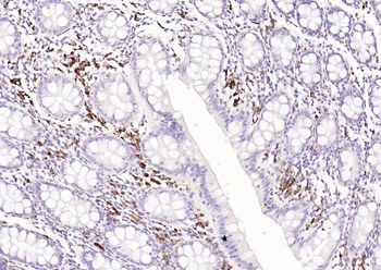
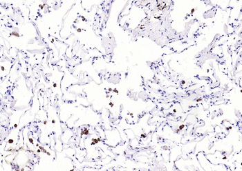
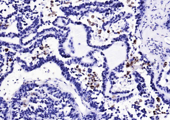
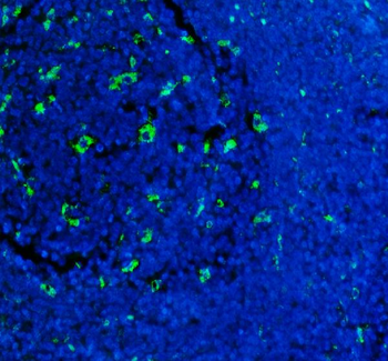
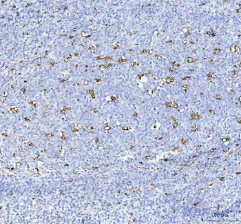
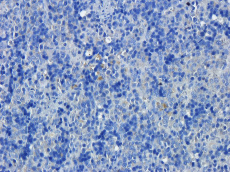
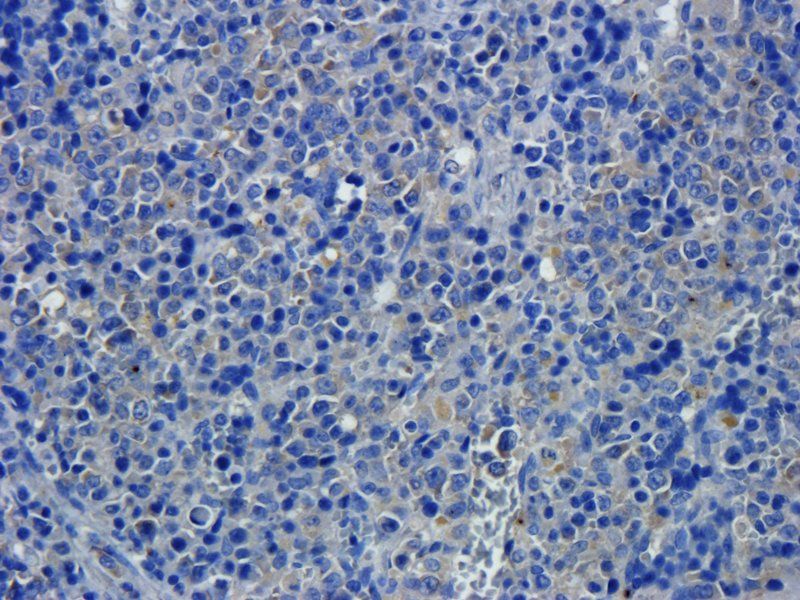
IHC-P image of mouse lymph node tissue using anti-CD68 (dilution of primary antibody at 1:100)
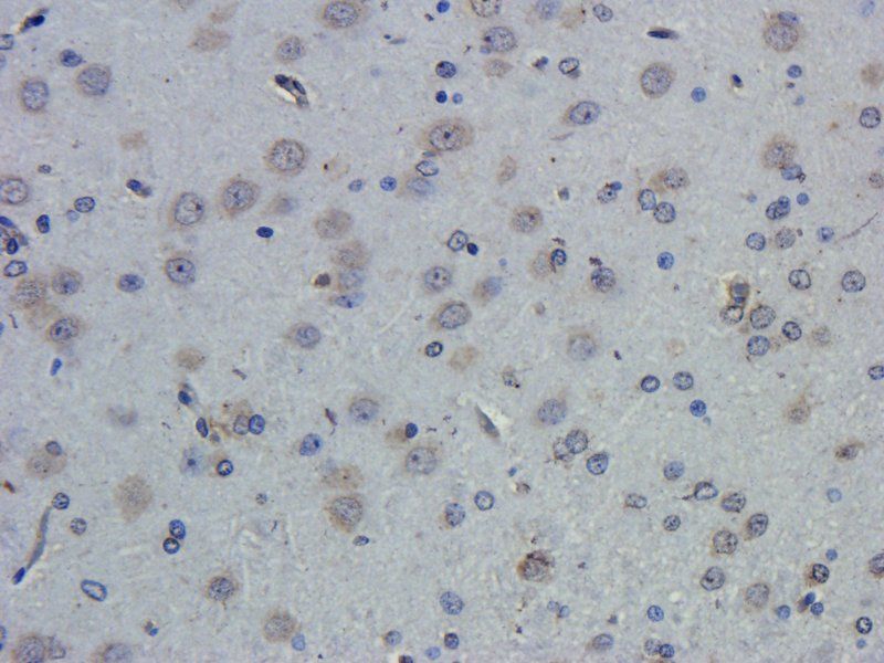
IHC-P staining of rat spleen tissue using anti-CD68 (dilution at 1:100)
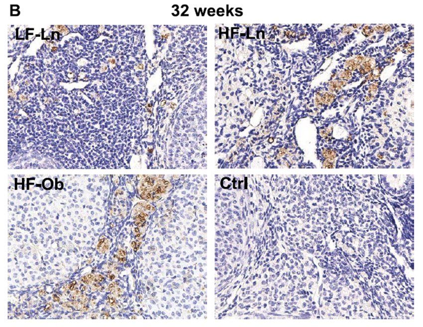
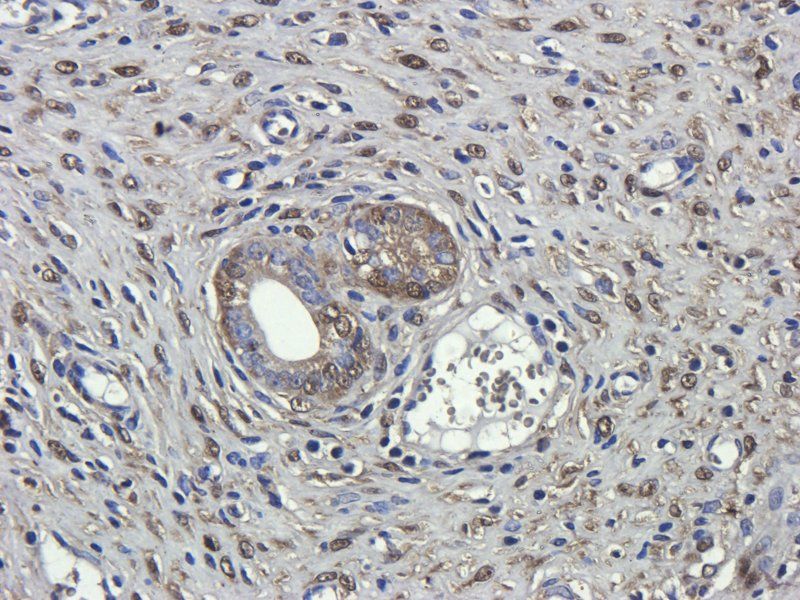
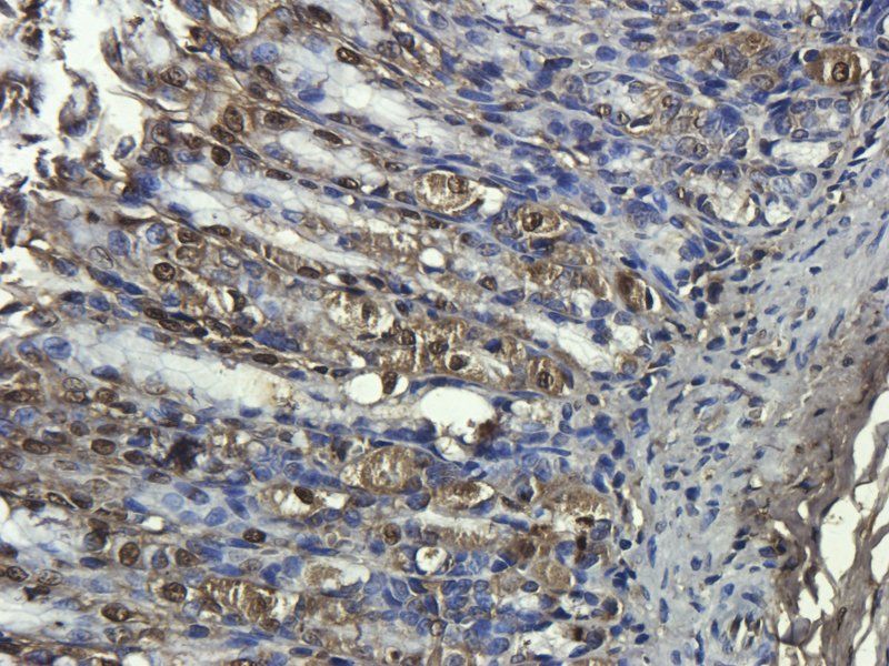
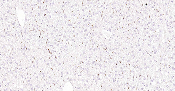
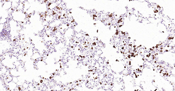
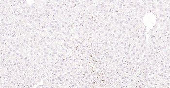
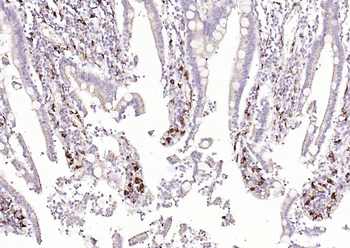
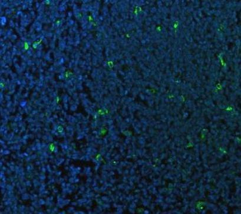
Representative images showing the presence of macrophages in the ovary. Macrophages were identified using marker CD68. Increased expression of macrophage marker CD68 was noted in both the HF-Ln (n¼6) and HF-Ob (n¼6) groups compared to the LF-Ln mice (n¼6)
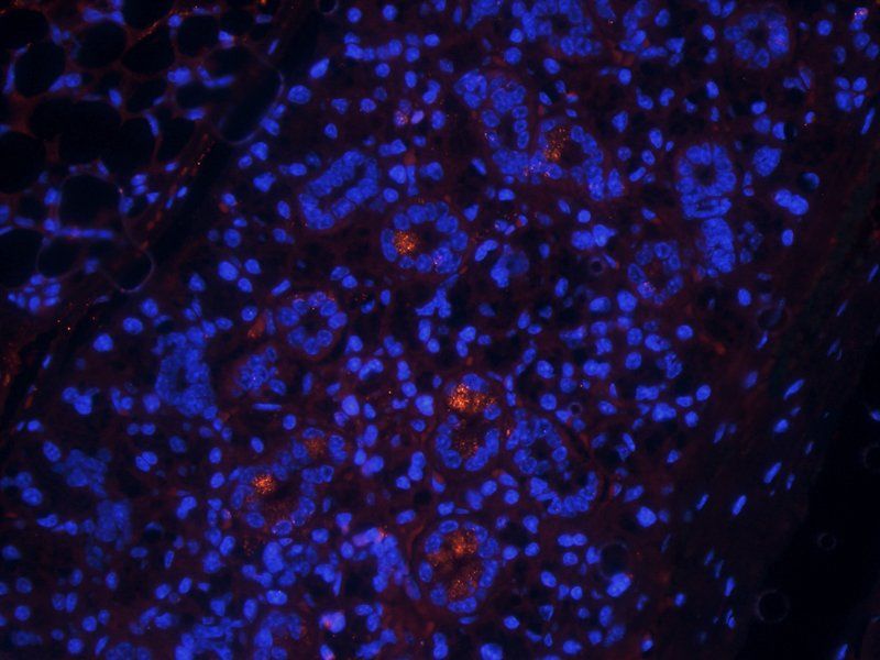
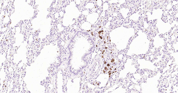
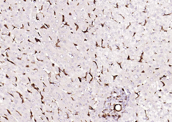
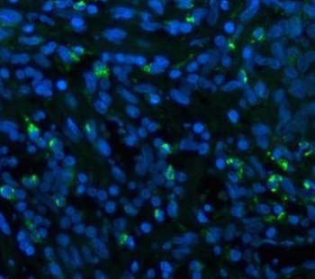
Immunofluorescence analysis of mouse lymph node tissue using CD68 antibody (dilution of primary antibody - 1:100)
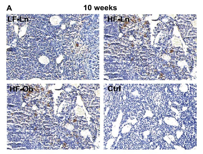
Representative images showing the presence of macrophages in the ovary. Macrophages were identified using marker CD68. Increased expression of macrophage marker CD68 was noted in both the HF-Ln (n¼6) and HF-Ob (n¼6) groups compared to the LF-Ln mice (n¼6)
Quick Database Links
Protocol Information
WB
Western Blot (IB, immunoblot)
IHC-P
Immunohistochemistry Paraffin
IF
Immunofluorescence
ICC
Immunocytochemistry
ELISA
Enzyme-linked Immunosorbent Assay (EIA)
Filter by Applications
Filter by Species
Skaznik-Wikiel, Malgorzata E. et al. High-Fat Diet Causes Subfertility and Compromised Ovarian Function Independent of Obesity in Mice Biol Reprod, 94, 108 (2016)
Applications
IHC
Reactivity
Mouse
Catarina Barbosa-Matos 1 2, Caroline Borges-Pereira 1 2, Sofia Libório-Ramos 1 2, Raquel Fernandes 1 2, Marcela Oliveira 1 2, Ana Mendes-Frias 1 2, Ricardo Silvestre 1 2, Nuno S Osório 1 2, Hélder N Bastos 3 4 5, Rita F Santos 4 6, Susana Guimarães 7, António Morais 3, Massimiliano Mazzone 8 9, Agostinho Carvalho 1 2, Cristina Cunha 1 2, Sandra Costa Deregulated immune cell recruitment orchestrated by c-MET impairs pulmonary inflammation and fibrosis Respir Res, 25, 257 (2024)
Applications
IF
Reactivity
Human
Yen-Zhen Lu, Ching-Ying Huang, Yi-Cheng Huang, Tsung-Chun Lee, Wei-Ting Kuo, Yu-Chen Pai, Linda Chia-Hui Yu Tumor Necrosis Factor α-Dependent Neutrophil Priming Prevents Intestinal Ischemia/Reperfusion-Induced Bacterial Translocation Digestive Diseases and Sciences, 62, 6 (2017)
Applications
IF
Reactivity
Rat
Wei-Shan Hsieh, Chia-Chi Kung, Shir-Ly Huang, Shih-Chang Lin, Wei-Hsin Sun TDAG8, TRPV1, and ASIC3 involved in establishing hyperalgesic priming in experimental rheumatoid arthritis Scientific Reports, 7, 8870 (2017)
Applications
IHC
Reactivity
Mouse
Jelodari, Sahar et al. Assessment of the Efficacy of an LL-37-Encapsulated Keratin Hydrogel for the Treatment of Full-Thickness Wounds ACS Appl Bio Mater, (2023)
CD68 Rabbit Polyclonal Antibody (orb197999)

- 0.0
Based on 0 reviews
Participating in our Biorbyt product reviews program enables you to support fellow scientists by sharing your firsthand experience with our products.
Login to Submit a ReviewAvailable Sizes
Select a size below
Choose Conjugation or Carrier Free Version
Free Secondary Antibody (20 ul)0/0
Please add an antibody product to your cart first.