You have no items in your shopping cart.
Featured
Description
Research Area
Immunology & Inflammation
Images & Validation
−Item 1 of 17
| Tested Applications | ELISA, ICC, IF, IHC-P, WB |
|---|---|
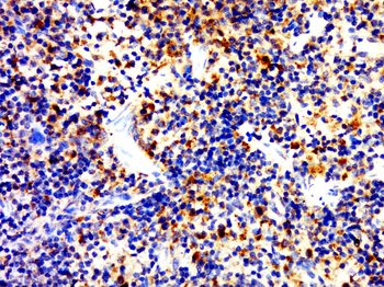
| Dilution Range | IHC-P: 1:500, IF/ICC: 1:200 |
| Reactivity | Human, Mouse, Rat |
Key Properties
−| Host | Rabbit |
|---|---|
| Clonality | Polyclonal |
| Isotype | IgG |
| Immunogen | KLH conjugated synthetic peptide derived from human CD4. Please contact us for the exact immunogen sequence. The peptide is available as orb374775. |
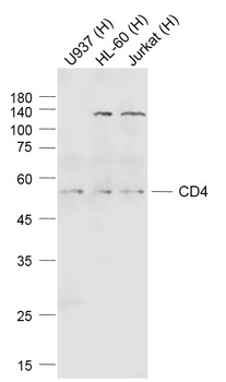
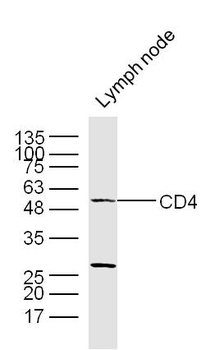
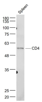
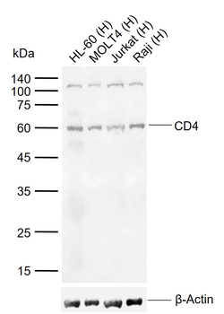
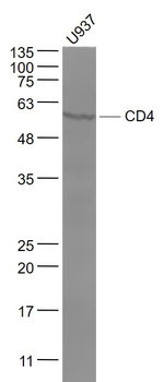
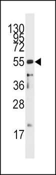
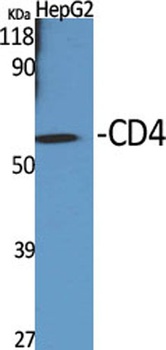
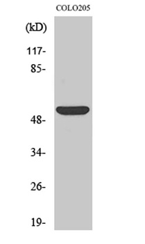
| Target | CD4 |
| Molecular Weight | 51 kDa |
| Purity | Polyclonal antibodies are purified by peptide affinity chromatography |
| Conjugation | Unconjugated |
Storage & Handling
−| Storage | Maintain refrigerated at 2-8°C for up to 2 weeks. For long term storage store at -20°C in small aliquots to prevent freeze-thaw cycles. |
|---|---|
| Form/Appearance | 10 mM PBS, 0.02% sodium azide |
| Concentration | - 100 μg (in 200 μl): 0.5 mg/ml- 200 μg (in 400 μl): 0.5 mg/ml |
| Expiration Date | 12 months from date of receipt. |
| Disclaimer | For research use only |
Alternative Names
−Anti-CD 4 antibody, Anti-CD4 (L3T4) antibody, Anti-CD4 antibody, Anti-CD4 antigen (p55) antibody, Anti-CD4 antigen antibody, Anti-CD4 molecule antibody, Anti-CD4 receptor antibody, Anti-CD4+ Lymphocyte deficiency, included antibody, Anti-CD4_HUMAN antibody, Anti-CD4mut antibody, Anti-L3T4 antibody, Anti-Leu3 antibody, Anti-Ly-4 antibody, Anti-Lymphocyte antigen CD4 antibody, Anti-MGC165891 antibody, Anti-OTTHUMP00000238897 antibody, Anti-p55 antibody, Anti-T cell antigen T4 antibody, Anti-T cell antigen T4/LEU3 antibody, Anti-T cell differentiation antigen L3T4 antibody, Anti-T cell OKT4 deficiency, included antibody, Anti-T cell surface antigen T4/Leu 3 antibody, Anti-T cell surface antigen T4/Leu3 antibody, Anti-T cell surface glycoprotein CD4 antibody, Anti-T-cell surface antigen T4/Leu-3 antibody, Anti-T-cell surface glycoprotein CD4 antibody, Anti-W3/25 antibody, Anti-W3/25 antigen antibody
Similar Products
−- Item 1 of 7
CD4 Rabbit Polyclonal Antibody [orb182470]
FC, IF, IHC-Fr, IHC-P, WB
Bovine, Canine, Guinea pig, Rat, Sheep
Human
Rabbit
Polyclonal
Unconjugated
100 μl, 200 μl, 50 μl - Item 1 of 4
CD4 Rabbit Polyclonal Antibody [orb312176]
IF, IHC-Fr, IHC-P, WB
Human, Rat
Human, Mouse, Rat
Rabbit
Polyclonal
Unconjugated
50 μl, 100 μl, 200 μl - Item 1 of 5
CD4 Antibody (C-term) [orb2649321]
FC, IHC-P, WB
Monkey
Human, Mouse
Rabbit
Polyclonal
Unconjugated
50 μl, 100 μl - Item 1 of 4
- Item 1 of 3
CD4 (phospho Ser433) rabbit pAb Antibody [orb770862]
ELISA, IF, IHC
Human, Mouse
Polyclonal
Unconjugated
100 μl, 50 μl

Quality Guarantee
Explore bioreagents carefree to elevate your research. All our products are rigorously tested for performance. If a product does not perform as described on its datasheet, our scientific support team will provide expert troubleshooting, a prompt replacement, or a refund. For full details, please see our Terms & Conditions and Buying Guide. Contact us at [email protected].
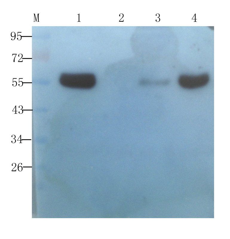
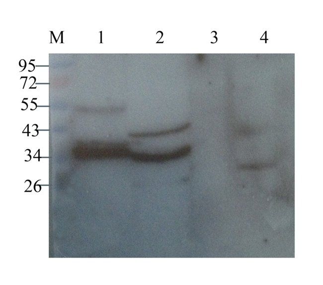
WB analysis of human breast cancer (lane 1), human thyroid cancer (lane 2), human endometrial cancer (lane 3), human ovarian cancer (lane 4) using CD4 antibody (1 ug/ml)
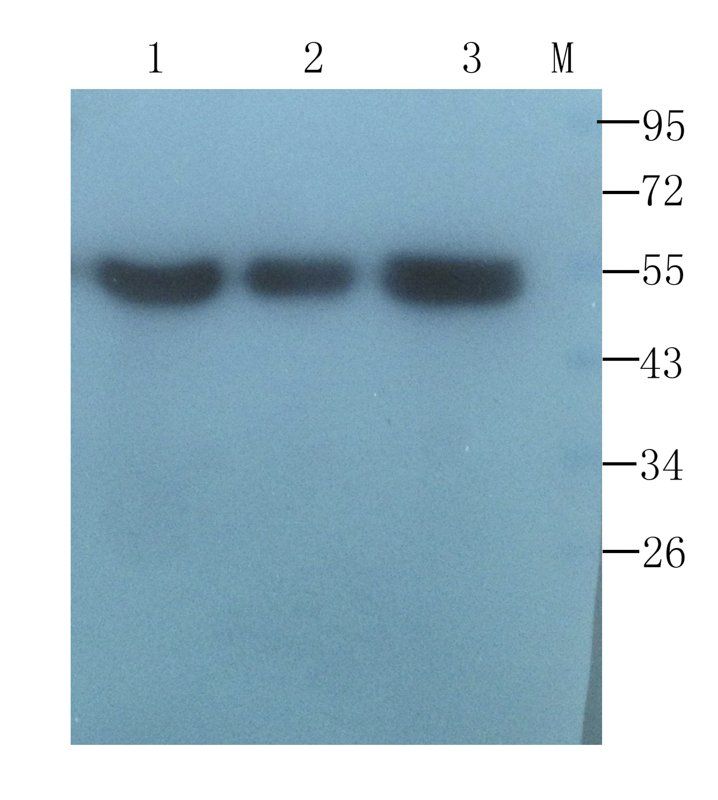
WB analysis of human breast tumor (lane 1), human mammary fibroma (lane 2), human breast cancer (lane 3) using CD4 antibody (1 ug/ml)
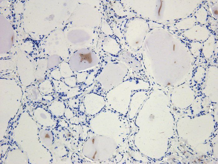
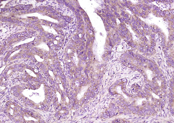
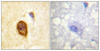
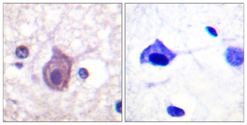
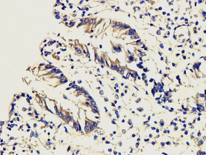
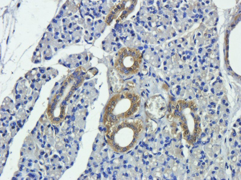
IHC-P image of human thyroid carcinoma tissue using CD4 antibody (2.5 ug/ml)
Western blot analysis of rat thymus (lane 1), rat liver (lane 2), mouse spleen (lane 3), mouse samll intestine (lane 4) using CD4 antibody (1 ug/ml)
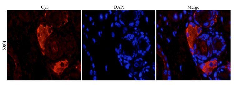
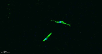
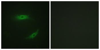
Immunofluorescence image of mouse skin tissue using CD4 antibody (2.5 ug/ml)
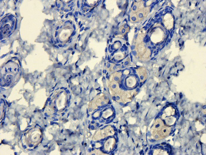
Immunohistochemical staining of paraffin embedded mouse skin tissue using CD4 antibody (2.5 ug/ml)
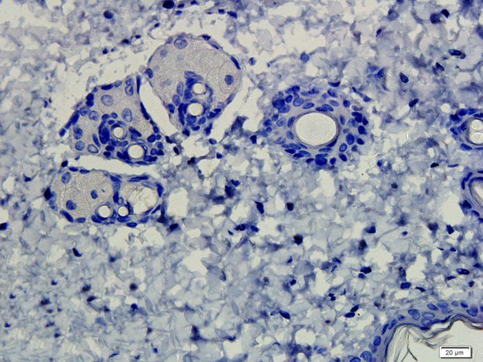
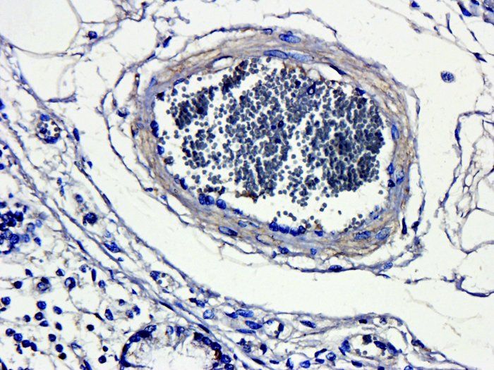
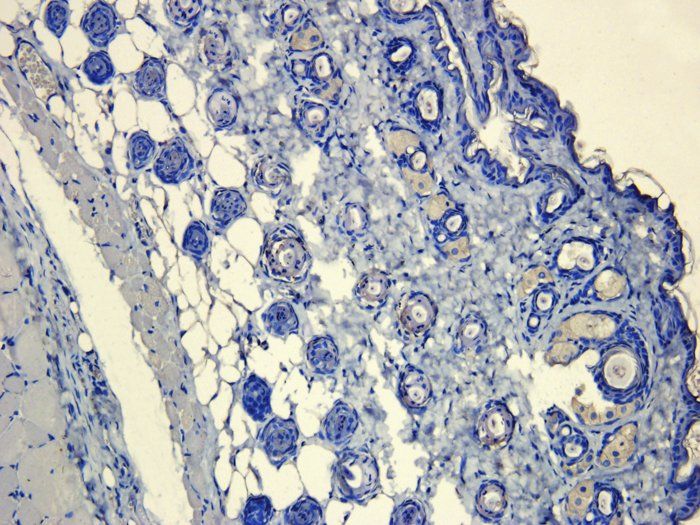
IHC-P image of rat skin tissue using CD4 antibody (2.5 ug/ml)
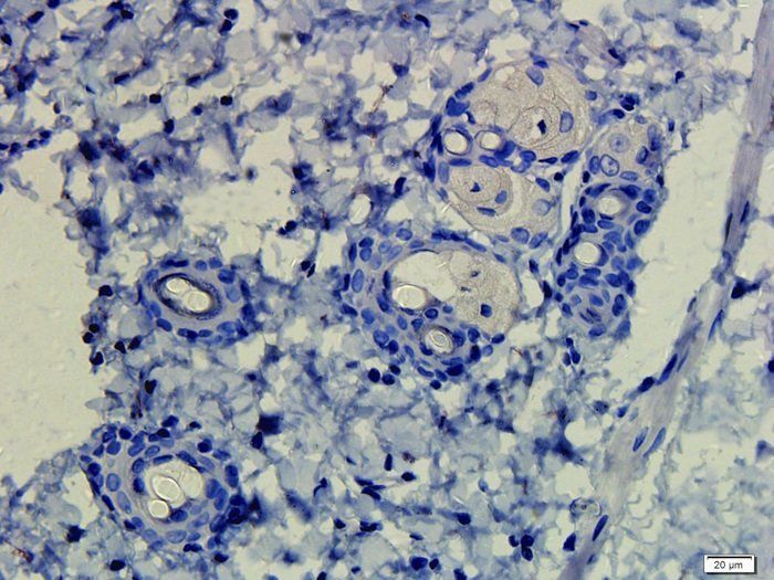
Immunohistochemical staining of rat skin tissue using anti-CD4 (2.5 ug/ml)
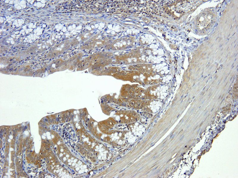
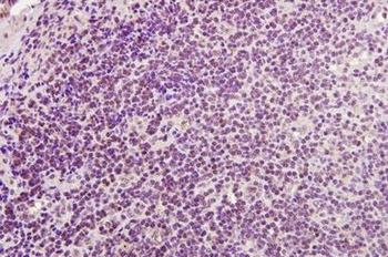
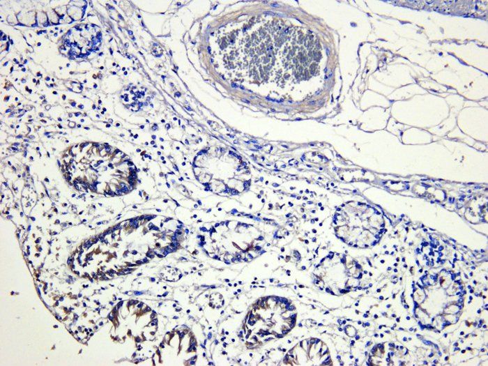
IHC-P staining of rat colon tissue using CD4 antibody (5 ug/ml)
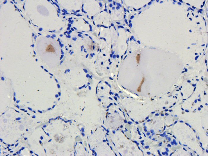
IHC-P staining of human thyroid carcinoma tissue using CD4 antibody (2.5 ug/ml)
Immunohistochemical staining of pig large intestines tissue using anti-CD4 (dilution of primary antibody - 1:200)
Immunohistochemical staining of paraffin embedded pig large intestines tissue using CD4 antibody (2.5 ug/ml)
IHC-P image of pig large intestines tissue using anti-CD4 (2.5 ug/ml)
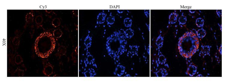
Immunofluorescence analysis of mouse skin tissue using CD4 antibody (2.5 ug/ml)
Immunohistochemical staining of paraffin embedded mouse skin tissue using CD4 antibody (2.5 ug/ml)
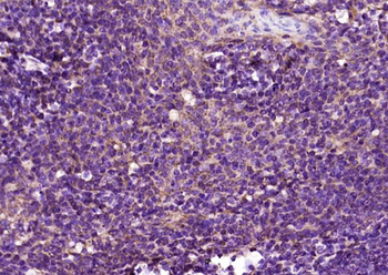
Immunohistochemical staining of mouse lymph node tissue using anti-CD4 (5 ug/ml)
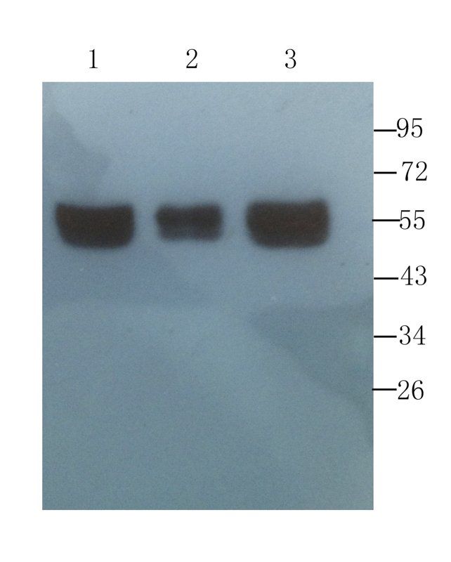
Western blot analysis of human breast tumor (lane 1), human mammary fibroma (lane 2), human breast cancer (lane 3) using CD4 antibody (1 ug/ml)
Quick Database Links
Protocol Information
WB
Western Blot (IB, immunoblot)
IHC-P
Immunohistochemistry Paraffin
IF
Immunofluorescence
ICC
Immunocytochemistry
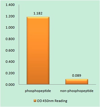
ELISA
Enzyme-linked Immunosorbent Assay (EIA)
Filter by Applications
Filter by Species
B. Sayyaf Dezfuli a, F. Pironi a, G. Castaldelli b, L. Giari b, M. Lanzoni b, K. Buchmann c, P.W. Kania c, G. Bosi Anguilla anguilla vs Contracaecum rudolphii: Granuloma allows host tolerance and parasite survival Aquaculture, (2024)
Applications
IHC
Mohammad Jahan-Mahin 1, Roya Askari 1, Amir Hossein Haghighi 1, Omid Khaiyat The effect of three types of water-based training protocols on thymus atrophy and specific indicators of cellular immune senescence in aged male rats Biogerontology, (2025)
Applications
IHC
Reactivity
Rat
Urszula Daniluk # 1, Agnieszka Świdnicka-Siergiejko # 2, Jarosław Daniluk 2, Małgorzata Rusak 3, Milena Dąbrowska 3, Katarzyna Guzińska-Ustymowicz 4, Anna Pryczynicz 4, Andrzej Dąbrowski 2 The development of pancreatic cancer is accompanied by significant changes in the immune response in genetically predisposed mice Front Oncol ., (2025)
Applications
IHC
Reactivity
Mouse
Romero-Moreno, Ricardo et al. The CXCL5/CXCR2 axis is sufficient to promote breast cancer colonization during bone metastasis Nat Commun, 10, 4404 (2019)
Applications
IHC
Reactivity
Mouse
Su, Jiyan et al. Protective Effect of Pogostone on 2,4,6-Trinitrobenzenesulfonic Acid-Induced Experimental Colitis via Inhibition of T Helper Cell Front Pharmacol, 8, 829 (2017)
Applications
IHC
Reactivity
Rat
CD4 Rabbit Polyclonal Antibody (orb4830)

- 0.0
Based on 0 reviews
Participating in our Biorbyt product reviews program enables you to support fellow scientists by sharing your firsthand experience with our products.
Login to Submit a ReviewAvailable Sizes
Select a size below
Choose Conjugation or Carrier Free Version
Free Secondary Antibody (20 ul)0/0
Please add an antibody product to your cart first.