You have no items in your shopping cart.
Description
Research Area
Metabolism Research
Images & Validation
−Item 1 of 3
| Tested Applications | ELISA, IHC, IP, WB |
|---|---|
| Dilution Range | ELISA: 1:10,000 - 1:50,000, IHC: 1:500 - 1:3,000, WB: 1:500 - 1:3,000 |
| Reactivity | Human |
| Application Notes |
Key Properties
−| Antibody Type | Primary Antibody |
|---|---|
| Host | Rabbit |
| Clonality | Polyclonal |
| Isotype | IgG |
| Immunogen | This affinity-purified antibody was prepared from whole rabbit serum produced by repeated immunizations with a synthetic peptide corresponding to the C-Terminal portion of Human Cbl-c. |
| Target | CBLC |
| Purity | This affinity purified antibody is directed against human Cbl-c protein. The product was affinity purified from monospecific antiserum by immunoaffinity purification. A BLAST analysis was used to suggest that this antibody would react with Cbl-c from human and chimpanzee sources. Expect partial reactivity against mouse and rat sources of Cbl-c as ~83% sequence homology is on record for the immunogen sequence. Reactivity with Cbl-c from other sources has not been determined. No reactivity is expected with Cbl-a or Cbl-b. |
| Conjugation | Unconjugated |
Storage & Handling
−| Storage | Store vial at -20° C prior to opening. Aliquot contents and freeze at -20° C or below for extended storage. Avoid cycles of freezing and thawing. Centrifuge product if not completely clear after standing at room temperature. This product is stable for several weeks at 4° C as an undiluted liquid. Dilute only prior to immediate use. |
|---|---|
| Form/Appearance | Liquid (sterile filtered) |
| Buffer/Preservatives | Preservative: 0.01% (w/v) Sodium Azide. Stabilizer: None; Buffer: 0.02 M Potassium Phosphate, 0.15 M Sodium Chloride, pH 7.2 |
| Concentration | 1.2 mg/ml |
| Expiration Date | 12 months from date of receipt. |
| Dry Ice Shipping | Please note: This product requires shipment on dry ice. A dry ice surcharge will apply. |
| Disclaimer | For research use only |
Alternative Names
−rabbit anti-Cbl-c Antibody, E3 ubiquitin-protein ligase CBL-C, RING finger protein 57, RING-type E3 ubiquitin transferase CBL-C, SH3-binding protein CBL-3, SH3-binding protein CBL-C, Signal transduction protein CBL-C, CBLC, CBL3, RNF57
Similar Products
−- Item 1 of 5
MMACHC Rabbit Polyclonal Antibody [orb556388]
ICC, IHC-P, WB
Human, Mouse, Rat
Rabbit
Polyclonal
Unconjugated
100 μl - Item 1 of 3
- Item 1 of 3
- Item 1 of 2
- Item 1 of 2


Quality Guarantee
Explore bioreagents carefree to elevate your research. All our products are rigorously tested for performance. If a product does not perform as described on its datasheet, our scientific support team will provide expert troubleshooting, a prompt replacement, or a refund. For full details, please see our Terms & Conditions and Buying Guide. Contact us at [email protected].
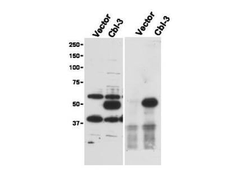
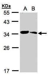
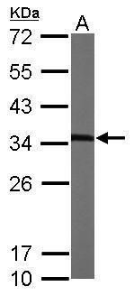
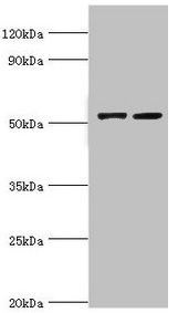
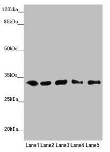
Immunoprecipitation (right) and western blot (left) using Biorbyt's Affinity Purified anti-Cbl-c antibody shows detection of a predominant band at ~52 kDa corresponding to Cbl-c. Lysates used are from HEK293T cells transfected with empty vector or with Cbl-c and western blotting (left panel). The predicted size of Cbl-c is 52 kDa. Size markers in kDa are shown to the left of the panel. The (right panel) shows immunoprecipitation with Rabbit anti-Cbl-c followed by western blotting using a Goat anti-Cbl-c antibody.
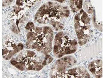
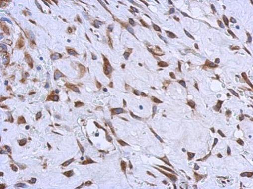
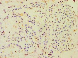
Biorbyt's Affinity Purified anti-Cbl-c antibody shows strong nuclear and cytoplasmic staining of cells in tubuli in human kidney tissue. Tissue was formalin-fixed and paraffin embedded. Brown color indicates presence of protein, blue color shows cell nuclei.
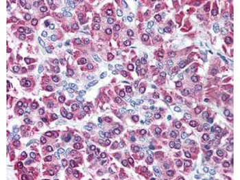
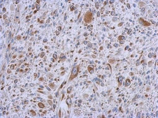
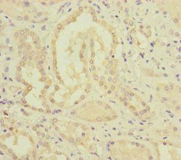
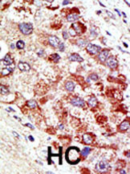
Biorbyt's affinity purified anti-Cbl-c antibody was used at 5 µg/ml to detect signal in a variety of tissues including multi-human, multi-brain and multi-cancer slides. This image shows moderate intracellular positive staining of human pancreatic acinar epithelium at 40X. Tissue was formalin-fixed and paraffin embedded. The image shows localization of the antibody as the precipitated red signal, with a hematoxylin purple nuclear counterstain.
Documents Download
Datasheet
Product Information
Request a Document
Protocol Information
WB
Western Blot (IB, immunoblot)
IHC
Immunohistochemistry
ELISA
Enzyme-linked Immunosorbent Assay (EIA)
IP
Immunoprecipitation
CBLC Antibody (orb345519)

- 0.0
Based on 0 reviews
Participating in our Biorbyt product reviews program enables you to support fellow scientists by sharing your firsthand experience with our products.
Login to Submit a ReviewAvailable Sizes
Select a size below
Choose Conjugation or Carrier Free Version
Free Secondary Antibody (20 ul)0/0
Please add an antibody product to your cart first.