You have no items in your shopping cart.
Featured
Description
Research Area
Cancer Biology, Neuroscience, Pharmacology & Drug Discovery, Signal Transduction
Images & Validation
−Item 1 of 4
| Tested Applications | AM, ICC, IF, IHC, IP, WB |
|---|---|
| Dilution Range | WB (1:1000), IHC-P (1:1000), ICC/IF (1:100) |
| Reactivity | Human, Mouse, Rat |
| Application Notes |
Key Properties
−| Host | Mouse |
|---|---|
| Clonality | Monoclonal |
| Isotype | IgG1 |
| Clone No. | N55/10 (Formerly sold as S55-10) |
| Immunogen | Fusion protein amino acids 1019-1293 (II-III loop) of human Cav3.2 |
| Target | Cav3.2 |
| Molecular Weight | 260kDa |
| Purification | Protein G Purified |
| Conjugation | Unconjugated |
Storage & Handling
−| Storage | Maintain refrigerated at 2-8°C for up to 2 weeks. For long term storage store at -20°C in small aliquots to prevent freeze-thaw cycles. |
|---|---|
| Buffer/Preservatives | PBS pH 7.4, 50% glycerol, 0.09% sodium azide. Storage buffer changes when conjugated. |
| Concentration | 1 mg/ml |
| Expiration Date | 12 months from date of receipt. |
| Disclaimer | For research use only |
Alternative Names
−Voltage-dependent T-type calcium channel subunit alpha-1H, Voltage-gated calcium channel subunit alpha Cav3.2, CACNA1H, KIAA1120, Cav3.2, CACNA1HB, calcium channel, voltage-dependent, T type, alpha 1H subunit, alpha 1Hb subunit, ECA6, EIG6, FLJ90484, Low-voltage-activated calcium channel alpha1 3.2 subunit, low-voltage-activated calcium channel alpha13.2 subunit, voltage dependent t-type calcium channel alpha-1H subunit, voltage-dependent T-type calcium channel subunit alpha-1H, voltage-gated calcium channel alpha subunit Cav3.2, voltage-gated calcium channel alpha subunit CavT.2
Similar Products
−- Item 1 of 1
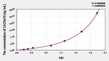
Mouse Calcium Channel, Voltage Dependent, T-Type, Alpha 1H Subunit (CACNa1H) ELISA Kit [orb780719]
Mouse
0.32-20 ng/mL
0.121 ng/mL
96 T, 48 T - Item 1 of 2
- Item 1 of 4
Cav3.2 Antibody (APC) [orb148211]
AM, ICC, IF, IHC, IP, WB
Human, Mouse, Rat
Mouse
Monoclonal
APC
100 μg - Item 1 of 4
Cav3.2 Antibody (Biotin) [orb148212]
AM, ICC, IF, IHC, IP, WB
Human, Mouse, Rat
Mouse
Monoclonal
Biotin
100 μg - Item 1 of 4
Cav3.2 Antibody (FITC) [orb148213]
AM, ICC, IF, IHC, IP, WB
Human, Mouse, Rat
Mouse
Monoclonal
FITC
100 μg


Quality Guarantee
Explore bioreagents carefree to elevate your research. All our products are rigorously tested for performance. If a product does not perform as described on its datasheet, our scientific support team will provide expert troubleshooting, a prompt replacement, or a refund. For full details, please see our Terms & Conditions and Buying Guide. Contact us at [email protected].

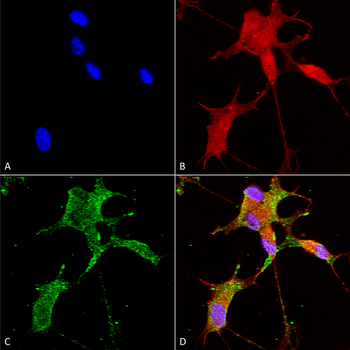
Immunocytochemistry/Immunofluorescence analysis using Mouse Anti-Cav3.2 Monoclonal Antibody, Clone N55/10. Tissue: Neuroblastoma cells (SH-SY5Y). Species: Human. Fixation: 4% PFA for 15 min. Primary Antibody: Mouse Anti-Cav3.2 Monoclonal Antibody at 1:50 for overnight at 4°C with slow rocking. Secondary Antibody: AlexaFluor 488 at 1:1000 for 1 hour at RT. Counterstain: Phalloidin-iFluor 647 (red) F-Actin stain; Hoechst (blue) nuclear stain at 1:800, 1.6mM for 20 min at RT. (A) Hoechst (blue) nuclear stain. (B) Phalloidin-iFluor 647 (red) F-Actin stain. (C) Cav3.2 Antibody (D) Composite.

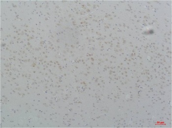
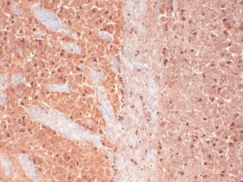
Immunohistochemistry analysis using Mouse Anti-CaV3.2 Calcium Channel Monoclonal Antibody, Clone N55/10. Tissue: hippocampus. Species: Human. Fixation: Bouin's Fixative and paraffin-embedded. Primary Antibody: Mouse Anti-CaV3.2 Calcium Channel Monoclonal Antibody at 1:1000 for 1 hour at RT. Secondary Antibody: FITC Goat Anti-Mouse (green) at 1:50 for 1 hour at RT.

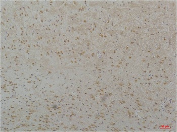
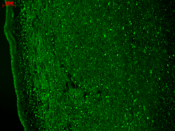
Immunohistochemistry analysis using Mouse Anti-CaV3.2 Calcium channel Monoclonal Antibody, Clone N55/10. Tissue: frozen brain section. Species: Human. Fixation: 10% Formalin Solution for 12-24 hours at RT. Primary Antibody: Mouse Anti-CaV3.2 Calcium channel Monoclonal Antibody at 1:1000 for 1 hour at RT. Secondary Antibody: HRP/DAB Detection System: Biotinylated Goat Anti-Mouse, Streptavidin Peroxidase, DAB Chromogen (brown) for 30 minutes at RT. Counterstain: Mayer Hematoxylin (purple/blue) nuclear stain at 250-500 μl for 5 minutes at RT.

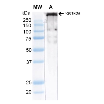
Western Blot analysis of Rat brain membrane lysate (native) showing detection of ~261 kDa Cav3.2 protein using Mouse Anti-Cav3.2 Monoclonal Antibody, Clone N55/10. Block: 2% Skim Milk + 2% BSA in TBST. Primary Antibody: Mouse Anti-Cav3.2 Monoclonal Antibody at 1:1000 for 2 hours at RT. Secondary Antibody: Anti-Mouse: HRP at 1:4000. Predicted/Observed Size: ~261 kDa.
Quick Database Links
UniProt Details
− No UniProt data available
NCBI Gene Details
− No NCBI Gene data available
NCBI Reference Sequences
−Associated Accession Numbers
Curated reference sequences for the gene transcript and protein product| Protein | NP_001005407.1 |
|---|
Documents Download
Datasheet
Product Information
Request a Document
Protocol Information
WB
Western Blot (IB, immunoblot)
IHC
Immunohistochemistry
IF
Immunofluorescence
ICC
Immunocytochemistry
IP
Immunoprecipitation
Cav3.2 Antibody (orb67393)

- 0.0
Based on 0 reviews
Participating in our Biorbyt product reviews program enables you to support fellow scientists by sharing your firsthand experience with our products.
Login to Submit a ReviewAvailable Sizes
Select a size below
Choose Conjugation or Carrier Free Version
Free Secondary Antibody (20 ul)0/0
Please add an antibody product to your cart first.