You have no items in your shopping cart.
Description
Research Area
Stem Cell & Developmental Biology
Images & Validation
−Item 1 of 7
| Tested Applications | ELISA, WB |
|---|---|
| Dilution Range | ELISA: 1:45,000, WB: 1:1,000 - 1:5,000 |
| Reactivity | Human |
| Application Notes |
Key Properties
−| Antibody Type | Primary Antibody |
|---|---|
| Host | Rabbit |
| Clonality | Polyclonal |
| Isotype | IgG |
| Immunogen | This affinity purified antibody was prepared from whole rabbit serum produced by repeated immunizations with a synthetic peptide corresponding to an internal region of Human Casz1 protein. |
| Target | CASZ1 |
| Purity | This affinity purified antibody is directed against human CASZ1 protein. The product was affinity purified from monospecific antiserum by immunoaffinity purification. A BLAST analysis was used to suggest reactivity with CASZ1 proteins from human, mouse, Drosophila, chimpanzee, and macaque based on a 100% homology. Partial reactivity is expected with horse and dog CASZ1 based on a 92% homology with the immunizing sequence. Cross-reactivity with CASZ1 from other sources has not been determined. |
| Conjugation | Unconjugated |
Storage & Handling
−| Storage | Store vial at -20° C prior to opening. Aliquot contents and freeze at -20° C or below for extended storage. Avoid cycles of freezing and thawing. Centrifuge product if not completely clear after standing at room temperature. This product is stable for several weeks at 4° C as an undiluted liquid. Dilute only prior to immediate use. |
|---|---|
| Form/Appearance | Liquid (sterile filtered) |
| Buffer/Preservatives | Preservative: 0.01% (w/v) Sodium Azide. Stabilizer: None; Buffer: 0.02 M Potassium Phosphate, 0.15 M Sodium Chloride, pH 7.2 |
| Concentration | 1.07 mg/mL |
| Expiration Date | 12 months from date of receipt. |
| Dry Ice Shipping | Please note: This product requires shipment on dry ice. A dry ice surcharge will apply. |
| Disclaimer | For research use only |
Alternative Names
−rabbit anti-CASZ1 Antibody, CASZ1, Zinc finger protein castor homolog 1, Castor-related protein, Zinc finger protein 693, CST, SRG, ZNF693
Similar Products
−- Item 1 of 7
- Item 1 of 4
ZNF693 Rabbit Polyclonal Antibody [orb101244]
IF, IHC-Fr, IHC-P
Human
Mouse, Rat
Rabbit
Polyclonal
Unconjugated
50 μl, 100 μl, 200 μl - Item 1 of 2
- Item 1 of 1
- Item 1 of 2
CASZ1 Rabbit Polyclonal Antibody [orb574530]
WB
Bovine, Equine, Guinea pig, Human, Rat
Mouse
Rabbit
Polyclonal
Unconjugated
100 μl


Quality Guarantee
Explore bioreagents carefree to elevate your research. All our products are rigorously tested for performance. If a product does not perform as described on its datasheet, our scientific support team will provide expert troubleshooting, a prompt replacement, or a refund. For full details, please see our Terms & Conditions and Buying Guide. Contact us at [email protected].
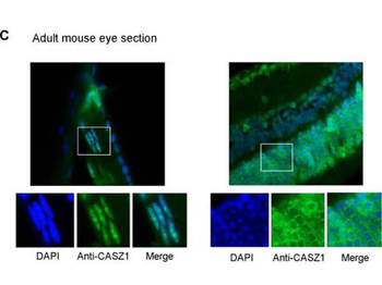
Immunofluorescence of Rabbit anti-CASZ1 Antibody. Tissue: adult murine ocular tissue. Antibody: Rabbit Anti-CASZ1 Antibody. Counterstain: DAPI. Localization: nucleus in lens epithelia but primarily localizes in the cytoplasm in photoreceptor cells.

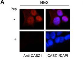
Immunofluorescence results of Endogenous CASZ1. Cells: BE2 cells. With or without Pre-Incubation of Anti-CASZ1 Antibody with CASZ1 Peptide. Staining: Rabbit Anti-CASZ1 Antibody. Chromatin counter stain: DAPI.
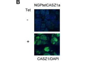
Immunofluorescence results of Rabbit Anti-CASZ1 Antibody. Tissue: Mouse Xenograft tumor of human NB cell line transfected with or without tetracycline inducible CASZ1 (NGPtetCASZ1a). Antibody: Rabbit Anti-CASZ1 Antibody. Counterstain: DAPI.

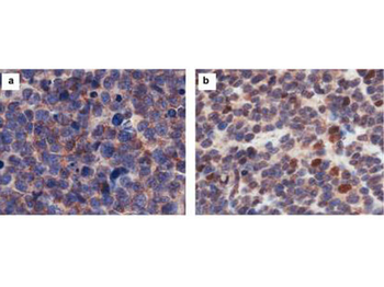
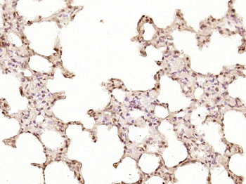
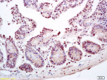
Immunohistochemistry results of Rabbit Anti-hCasz1 Antibody. Tissue: NB patient tumor. A. CASZ1 localized exclusively in the cytoplasm. B. CASZ1 localized in the cytoplasm and nucleus. Primary Antibody: Rabbit Anti-CASZ1 stained brown. Nucleus counterstained with hematoxylin (blue).

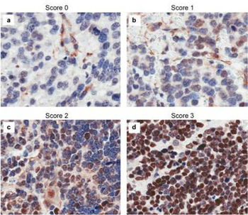
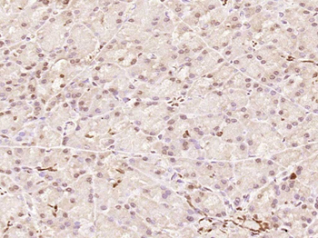
Immunohistochemistry results of Rabbit Anti-hCASZ1 Antibody. Tissue: NB patient tumor. A. Score 0- a rare positive nuclei. B. Score 1- (1-10% positive) equivocal/uninterpretable. C. Score 2- (10-50% positive) weak positive. D. Score 3- (> 50% positive) strong positive. Primary Antibody: Rabbit Anti-CASZ1 stained brown. Nucleus counterstained with hematoxylin (blue). Localization: Nuclear.
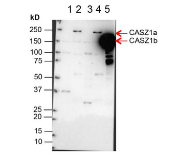
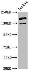
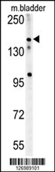
Western Blot of Anti-CASZ1 Antibody. Lane 1: NBLS Cytoplasmic (20 µg). Lane 2: NBLS Nuclear (3 µg). Lane 3: BE2C Cytoplasmic (30 µg). Lane 4: BE2C Nuclear (7 µg). Lane 5: SY5Y-CASZ1b (10 µg). Block: 5% Blotto/TTBS for 1 hour. Primary: Casz1 1:10000 for 1 hour. Secondary: Goat anti-Rabbit HRP for 1 hour. 240sec exposure. Detects nuclear endogenous CASZ1a and CASZ1b; and transiently transfected CASZ1b isoform.

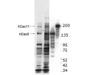
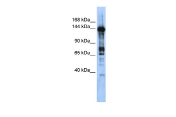
Western blot using Biorbyt's anti-hCASZ1 antibody. This blot shows detection of endogenous and transfected human CASZ1 protein in fresh whole cell lysate (~30 µg). Lane 1: BE2(s) cell lysate. Lane 2: BE2(N) cell lysate. Lane 3: SY5Y transfected with hCASZ5 (125kDa). Lane 4: SY5Y transfected with hCASZ11 (190kDa). Protein was resolved by SDS-PAGE and transferred onto nitrocellulose. After blocking, the membrane was probed with the primary antibody diluted to 1:1000 for 1.5 hours at room temperature then incubated with HRP-conjugated Goat Anti-Rabbit antibody for 45 min. at room temperature.
Documents Download
Datasheet
Product Information
Request a Document
Protocol Information
WB
Western Blot (IB, immunoblot)
ELISA
Enzyme-linked Immunosorbent Assay (EIA)
CASZ1 Antibody (orb345675)

- 0.0
Based on 0 reviews
Participating in our Biorbyt product reviews program enables you to support fellow scientists by sharing your firsthand experience with our products.
Login to Submit a ReviewAvailable Sizes
Select a size below
Choose Conjugation or Carrier Free Version
Free Secondary Antibody (20 ul)0/0
Please add an antibody product to your cart first.