You have no items in your shopping cart.
Calbindin 1 antibody
SKU: orb2276324
Description
Images & Validation
−Item 1 of 3
| Tested Applications | IHC |
|---|---|
| Dilution Range | 1:50-1:100 |
| Reactivity | Human |
| Application Notes |
Key Properties
−| Host | Mouse |
|---|---|
| Clonality | Monoclonal |
| Isotype | IgG2c, kappa |
| Clone No. | MSVA-471M |
| Immunogen | Recombinant fragment (around aa 7-96) of human CALB1 protein (exact sequence is proprietary) |
| Conjugation | Unconjugated |
Storage & Handling
−| Storage | Maintain refrigerated at 2-8°C for up to 2 weeks. For long term storage store at -20°C in small aliquots to prevent freeze-thaw cycles. |
|---|---|
| Expiration Date | 12 months from date of receipt. |
| Disclaimer | For research use only |
Alternative Names
−avian-type; CAB27; CALB 1; CALB; CALB1; CALB1_HUMAN; Calbindin 1 28kDa; Calbindin; Calbindin D28; D 28K; D-28K; D28K; OTTHUMP00000166027; OTTHUMP00000225441; RTVL H protein; Vitamin D dependent calcium binding protein; Vitamin D dependent calcium binding protein avian type; Vitamin D-dependent calcium-binding protein
Similar Products
−- Item 1 of 7
Calbindin/CALB1 Rabbit Polyclonal Antibody [orb443152]
ELISA, FC, IF, IHC, WB
Human, Mouse, Rat
Rabbit
Polyclonal
Unconjugated
100 μg - Item 1 of 4
calbindin D28 Antibody [orb12289]
ELISA, IHC, WB
Bovine, Canine, Porcine
Human, Rat
Goat
Polyclonal
Unconjugated
100 μg - Item 1 of 4
CALB1 Rabbit Polyclonal Antibody [orb625366]
ELISA, IF, IHC, IP, WB
Human, Mouse, Rat
Rabbit
Polyclonal
Unconjugated
50 μg, 100 μg - Item 1 of 3
- Item 1 of 3


Quality Guarantee
Explore bioreagents carefree to elevate your research. All our products are rigorously tested for performance. If a product does not perform as described on its datasheet, our scientific support team will provide expert troubleshooting, a prompt replacement, or a refund. For full details, please see our Terms & Conditions and Buying Guide. Contact us at [email protected].
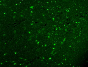
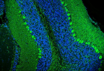
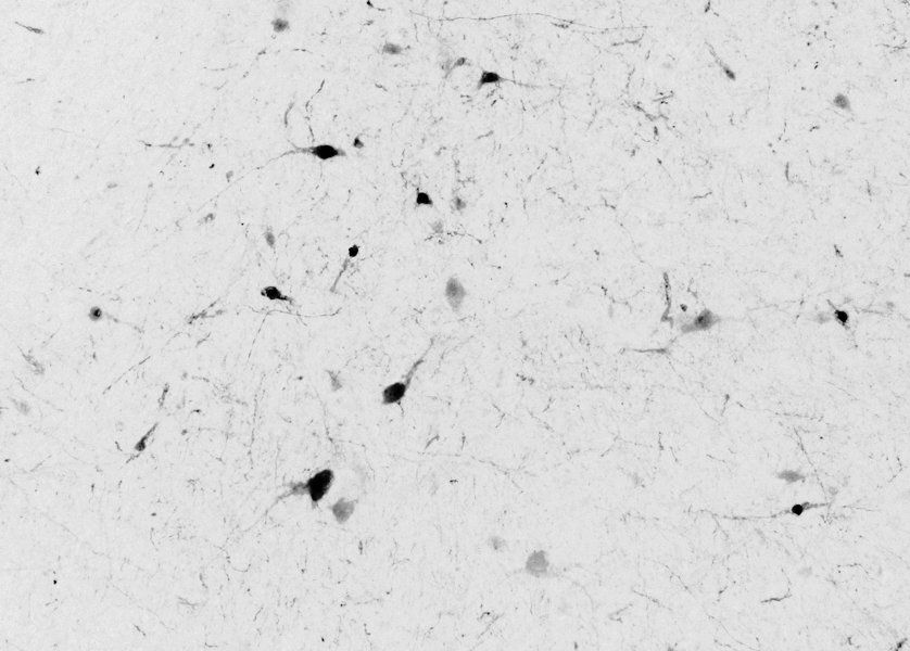
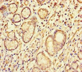
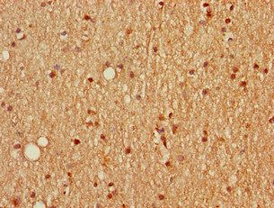
Cerebellum molecular layer Purkinje cell layer granule cell layer white matter A strong Calbindin 1 staining of Purkinje cells and of associated axonal fibres is seen in the cerebrum.
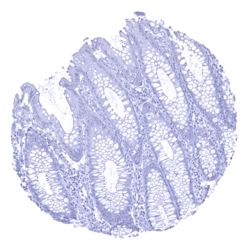
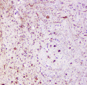
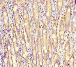
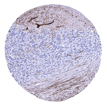
Colon descendes mucosa Calbindin 1 immunostaining is absent in all cells of the colon mucosa.
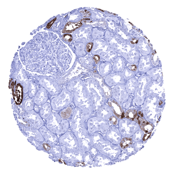
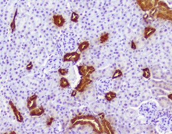
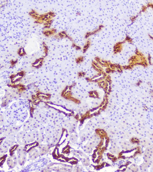
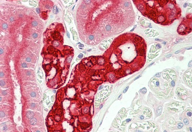
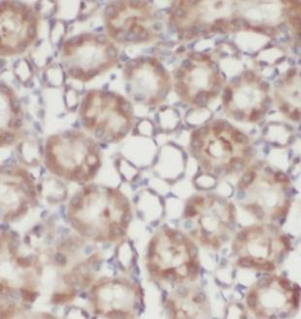
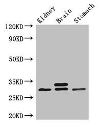
Kidney cortex A strong cytoplasmic Calbindin 1 immunostaining occurs in a fraction of distal tubuli of the kidney.
Quick Database Links
UniProt
UniProt Details
− No UniProt data available
Documents Download
Datasheet
Product Information
Request a Document
Calbindin 1 antibody (orb2276324)

- 0.0
Based on 0 reviews
Participating in our Biorbyt product reviews program enables you to support fellow scientists by sharing your firsthand experience with our products.
Login to Submit a ReviewAvailable Sizes
Select a size below