You have no items in your shopping cart.
Featured
Description
Research Area
Immunology & Inflammation
Images & Validation
−Item 1 of 8
| Tested Applications | ELISA, ICC, IF, IHC, WB |
|---|---|
| Reactivity | Human, Mouse, Rat |
Key Properties
−| Antibody Type | Primary Antibody |
|---|---|
| Host | Rabbit |
| Clonality | Polyclonal |
| Isotype | IgG |
| Immunogen | Anti-BAFF antibody (orb1239254) was raised against a peptide corresponding to 16 amino acids near the carboxy terminus of human BAFF. The immunogen is located within the last 50 amino acids of BAFF. |
| Target | TNFSF13B |
| Molecular Weight | Predicted: 31kDObserved: 35 kD |
| Purification | BAFF Antibody is Ion exchange chromatography purified. |
| Conjugation | Unconjugated |
Storage & Handling
−| Storage | Maintain refrigerated at 2-8°C for up to 2 weeks. For long term storage store at -20°C in small aliquots to prevent freeze-thaw cycles. |
|---|---|
| Form/Appearance | Liquid |
| Buffer/Preservatives | BAFF Antibody is supplied in PBS containing 0.02% sodium azide. |
| Concentration | 1 mg/mL |
| Expiration Date | 12 months from date of receipt. |
| Disclaimer | For research use only |
Alternative Names
−BAFF Antibody: DTL, BAFF, BLYS, CD257, TALL1, THANK, ZTNF4, TALL-1, TNFSF20, UNQ401/PRO738, Tumor necrosis factor ligand superfamily member 13B, B lymphocyte stimulator
Similar Products
−- Item 1 of 4
TALL-1 rabbit pAb Antibody [orb766771]
ELISA, IF, IHC, WB
Human, Mouse
Polyclonal
Unconjugated
50 μl, 100 μl - Item 1 of 1
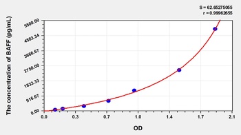
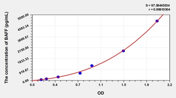
Human B-Cell Activating Factor (BAFF/CD257) ELISA Kit [orb776009]
Human
78.13-5000 pg/mL
28 pg/mL
48 T, 96 T - Item 1 of 1
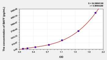
Rat B-Cell Activating Factor (BAFF/CD257) ELISA Kit [orb780111]
Rat
78.13-5000 pg/mL
26 pg/mL
48 T, 96 T - Item 1 of 1
Chicken B-Cell Activating Factor (BAFF/CD257) ELISA Kit [orb781644]
Gallus
78.13-5000 pg/mL
28 pg/mL
48 T, 96 T - Item 1 of 1
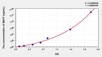
Pig B-Cell Activating Factor (BAFF/CD257) ELISA Kit [orb782106]
Porcine
0.16-10 ng/mL
0.057 ng/mL
48 T, 96 T

Quality Guarantee
Explore bioreagents carefree to elevate your research. All our products are rigorously tested for performance. If a product does not perform as described on its datasheet, our scientific support team will provide expert troubleshooting, a prompt replacement, or a refund. For full details, please see our Terms & Conditions and Buying Guide. Contact us at [email protected].
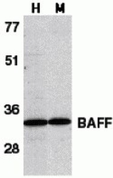
Western Blot Validation in Human HL60 Cell Lysate (H) and Mouse Spleen Lysate (M). Loading: 15 µg of lysates per lane. Antibodies: BAFF orb1239254 (1 µg/mL), 1h incubation at RT in 5% NFDM/TBST. Secondary: Goat anti-rabbit IgG HRP conjugate at 1:10000 dilution.
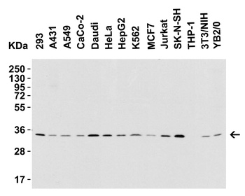
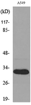
Western Blot Validation in Human, Mouse and Rat Cell Lines. Loading: 15 µg of lysates per lane. Antibodies: BAFF orb1239254 (1 µg/mL), 1h incubation at RT in 5% NFDM/TBST. Secondary: Goat anti-rabbit IgG HRP conjugate at 1:10000 dilution.
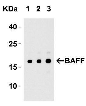
Western Blot Validation with Recombinant Protein. Loading: 30 ng of human BAFF recombinant protein per lane. Antibodies: BAFF orb1239254 (Lane 1: 0.25 µg/mL; Lane 2: 0.5 µg/mL and Lane 3: 1 µg/mL), 1h incubation at RT in 5% NFDM/TBST. Secondary: Goat anti-rabbit IgG HRP conjugate at 1:10000 dilution. Observed at around 18kD.
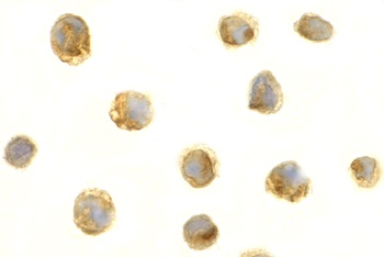
Immunocytochemistry Validation of BAFF in HL60 Cells. Immunocytochemical analysis of HL60 cells using anti-BAFF antibody (orb1239254) at 1 µg/ml. Cells was fixed with formaldehyde and blocked with 10% serum for 1 h at RT; antigen retrieval was by heat mediation with a citrate buffer (pH6). Samples were incubated with primary antibody overnight at 4°C. A goat anti-rabbit IgG H&L (HRP) at 1/250 was used as secondary. Counter stained with Hematoxylin.
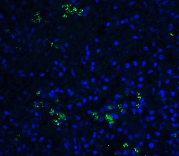
Immunofluorescence Validation of BAFF in Human Spleen Tissue. Immunofluorescent analysis of 4% paraformaldehyde-fixed human spleen tissue labeling BAFF with orb1239254 at 20 µg/mL, followed by goat anti-rabbit IgG secondary antibody at 1/500 dilution (green) and DAPI staining (blue).
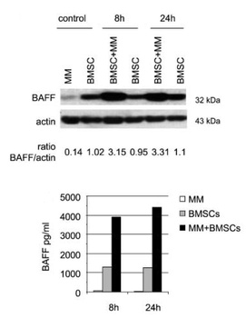
Regulated Expression Validation of BAFF in Myeloma Patients (Tai et al., 2006). Immunoblot analysis was performed to monitor protein expression of BAFF with anti-BAFF antibodies in multiple myeloma cells with or without BMSCs. BAFF expression in cocultures at 8hr or 24hr was up-regulated by ~3.5-fold relative to BMSCs alone.
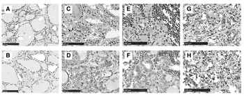
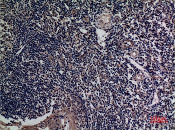
Immunohistochemistry Validation of BAFF in Thyroid of Patients with Graves' Diseases (Campi et al., 2015). BAFF expression detected by anti-BAFF antibodies (orb1239254) was remarkably increased in thyrocytes from multinodular goiter (C) compared with either Hashimoto's thyroiditis (E) or Graves' disease (G) while no staining was found in normal thyroid tissue (A).
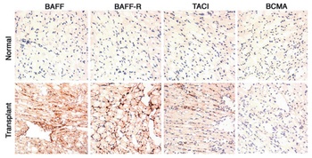
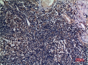
Immunohistochemistry Validation of BAFF in Murine Cardiac Transplants at Rejection (Ye et al., 2004). BAFF expression detected by anti-BAFF antibodies (orb1239254) was upregulated in intragraft leukocytes due to rejection at 7 days after heart transplant.
Documents Download
Datasheet
Product Information
Request a Document
Protocol Information
WB
Western Blot (IB, immunoblot)
IHC
Immunohistochemistry
IF
Immunofluorescence
ICC
Immunocytochemistry
ELISA
Enzyme-linked Immunosorbent Assay (EIA)
TNFSF13B Antibody (orb1239254)

- 0.0
Based on 0 reviews
Participating in our Biorbyt product reviews program enables you to support fellow scientists by sharing your firsthand experience with our products.
Login to Submit a ReviewAvailable Sizes
Select a size below
Free Secondary Antibody (20 ul)0/0
Please add an antibody product to your cart first.