You have no items in your shopping cart.
Description
Research Area
Signal Transduction
Images & Validation
−Item 1 of 7
| Tested Applications | FC, IF, IHC-P, WB |
|---|---|
| Dilution Range | IF - 1:25, WB - 1:2000, IHC-P - 1:10-50, FC - 1:25 |
| Reactivity | Human |
| Predicted Reactivity | Mouse |
Key Properties
−| Antibody Type | Primary Antibody |
|---|---|
| Host | Rabbit |
| Clonality | Polyclonal |
| Isotype | Rabbit IgG |
| Immunogen | This B-RAF antibody is generated from rabbits immunized with a KLH conjugated synthetic peptide between 424-453 amino acids from human B-RAF. Antigen Region: 424-453 aa. |
| Target | BRAF (HGNC:1097) |
| Molecular Weight | 84437 Da |
| Conjugation | Unconjugated |
Storage & Handling
−| Storage | Maintain refrigerated at 2-8°C for up to 2 weeks. For long term storage store at -20°C in small aliquots to prevent freeze-thaw cycles |
|---|---|
| Form/Appearance | Purified polyclonal antibody supplied in PBS with 0.09% (W/V) sodium azide. This antibody is purified through a protein A column, followed by peptide affinity purification. |
| Expiration Date | 12 months from date of receipt. |
| Disclaimer | For research use only |
Alternative Names
−Serine/threonine-protein kinase B-raf, Proto-oncogene B-Raf, p94, v-Raf murine sarcoma viral oncogene homolog B1, BRAF, BRAF1, RAFB1
Similar Products
−- Item 1 of 1

Phospho-B-Raf-S445 polyclonal antibody [orb642228]
WB
Human, Rat
Rabbit
Polyclonal
Unconjugated
200 μl, 100 μl, 50 μl
Quality Guarantee
Explore bioreagents carefree to elevate your research. All our products are rigorously tested for performance. If a product does not perform as described on its datasheet, our scientific support team will provide expert troubleshooting, a prompt replacement, or a refund. For full details, please see our Terms & Conditions and Buying Guide. Contact us at [email protected].
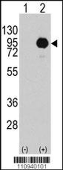
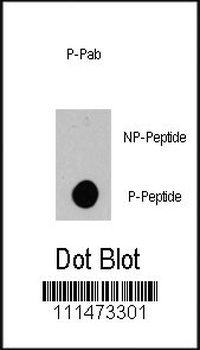
Western blot analysis of BRAF (arrow) using rabbit polyclonal BRAF Antibody (S445). 293 cell lysates (2 ug/lane) either nontransfected (Lane 1) or transiently transfected with the BRAF gene (Lane 2).
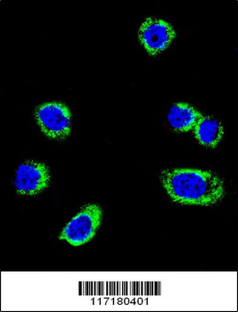
Confocal immunofluorescent analysis of B-RAF Antibody (S445) with Hela cell followed by Alexa Fluor 488-conjugated goat anti-rabbit lgG (green).DAPI was used to stain the cell nuclear (blue).
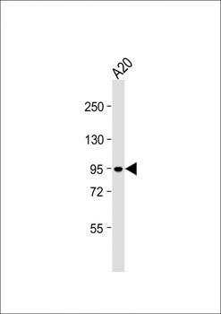
Anti-B-RAF Antibody (S445) at 1:2000 dilution + A20 whole cell lysate. Lysates/proteins at 20 µg per lane. Secondary Goat Anti-Rabbit IgG, (H+L), Peroxidase conjugated at 1/10000 dilution. Predicted band size: 84 kDa. Blocking/Dilution buffer: 5% NFDM/TBST.
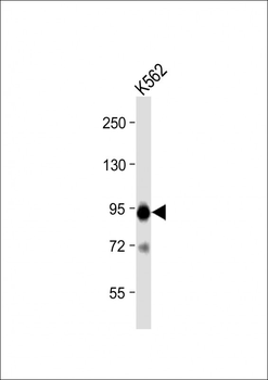
Anti-B-RAF Antibody (S445) at 1:2000 dilution + K562 whole cell lysate. Lysates/proteins at 20 µg per lane. Secondary Goat Anti-Rabbit IgG, (H+L), Peroxidase conjugated at 1/10000 dilution. Predicted band size: 84 kDa. Blocking/Dilution buffer: 5% NFDM/TBST.
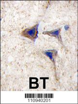
Formalin-fixed and paraffin-embedded human brain tissue reacted with BRAF Antibody (S445), which was peroxidase-conjugated to the secondary antibody, followed by DAB staining. This data demonstrates the use of this antibody for immunohistochemistry; clinical relevance has not been evaluated.
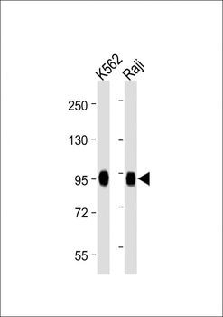
All lanes: Anti-B-RAF Antibody (S445) at 1:2000 dilution. Lane 1: K562 whole cell lysate. Lane 2: Raji whole cell lysate. Lysates/proteins at 20 µg per lane. Secondary Goat Anti-Rabbit IgG, (H+L), Peroxidase conjugated at 1/10000 dilution. Predicted band size: 84 kDa. Blocking/Dilution buffer: 5% NFDM/TBST.
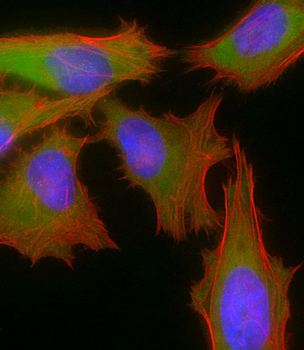
Immunofluorescent analysis of 4% paraformaldehyde-fixed, 0.1% Triton X-100 permeabilized HeLa (human cervical epithelial adenocarcinoma cell line) cells labeling B-RAF at 1/25 dilution, followed by Dylight 488-conjugated goat anti-rabbit IgG secondary antibody at 1/200 dilution (green). Immunofluorescence image showing cytoplasm and nucleus staining on HeLa cell line. Cytoplasmic actin is detected with Dylight 554 Phalloidin at 1/100 dilution (red). The nuclear counter stain is DAPI (blue).
Quick Database Links
UniProt Details
− No UniProt data available
NCBI Reference Sequences
−Associated Accession Numbers
Curated reference sequences for the gene transcript and protein product| Protein | NP_004324.2 |
|---|
Documents Download
Datasheet
Product Information
Request a Document
Protocol Information
WB
Western Blot (IB, immunoblot)
IHC-P
Immunohistochemistry Paraffin
FC
Flow Cytometry
IF
Immunofluorescence
B-RAF Antibody (S445) (orb1928866)

- 0.0
Based on 0 reviews
Participating in our Biorbyt product reviews program enables you to support fellow scientists by sharing your firsthand experience with our products.
Login to Submit a ReviewAvailable Sizes
Select a size below
Choose Conjugation or Carrier Free Version
Free Secondary Antibody (20 ul)0/0
Please add an antibody product to your cart first.