You have no items in your shopping cart.
ATG7 Antibody
SKU: orb54993
Description
Research Area
Cancer Biology, Cardiovascular Research, Cell Biology
Images & Validation
−Item 1 of 4
| Tested Applications | ICC, IF, IHC, WB |
|---|---|
| Dilution Range | WB (1:1000); ICC/IF (1:100); IHC-P (1:50) |
| Reactivity | Human, Mouse, Rat |
| Application Notes |
Key Properties
−| Host | Rabbit |
|---|---|
| Clonality | Polyclonal |
| Immunogen | Synthetic peptide from the N-terminal of Human ATG7 |
| Target | ATG7 |
| Molecular Weight | 77.9 kDa |
| Purification | Peptide Affinity Purified |
| Conjugation | Unconjugated |
Storage & Handling
−| Storage | Maintain refrigerated at 2-8°C for up to 2 weeks. For long term storage store at -20°C in small aliquots to prevent freeze-thaw cycles. |
|---|---|
| Buffer/Preservatives | PBS, 50% glycerol, 0.09% sodium azide Storage buffer changes when conjugated |
| Concentration | 1 mg/ml |
| Expiration Date | 12 months from date of receipt. |
| Disclaimer | For research use only |
Alternative Names
−ATG7, ATG7_HUMAN, APG7, APG7L, APG7-like, APG7 autophagy 7 like, APG7 autophagy 7-like (S. cerevisiae), ATG7 autophagy related 7 homolog, ATG7 autophagy related 7 homolog (S. cerevisiae), ATG12-activating enzyme E1 ATG7, Ubiquitin-like modifier-activating enzyme ATG7, Ubiquitin-activating enzyme E1-like protein, Ubiquitin activating enzyme E1 like protein, Autophagy-related protein 7, Autophagy related protein 7, Autophagy-related 7 (yeast), Autophagy 7 S. cerevisiae homolog of, GSA7, GSA 7, hAGP7, Atg7l, DKFZp434N0735, 1810013K23Rik, ATG 7, Apg 7
Similar Products
−- Item 1 of 8
ATG7 Rabbit Polyclonal Antibody [orb1819451]
ELISA, FC, ICC, IF, IHC, WB
Human
Rabbit
Polyclonal
Unconjugated
100 μg - Item 1 of 5
ATG7 Antibody (C-term) [orb1933432]
IF, IHC-P, WB
Gallus, Rat
Human, Mouse
Rabbit
Polyclonal
Unconjugated
100 μl, 50 μl - Item 1 of 6
ATG7 Rabbit Polyclonal Antibody [orb654295]
FC, IHC, WB
Human, Mouse, Rat
Rabbit
Polyclonal
Unconjugated
100 μg - Item 1 of 4
- Item 1 of 4
ATG7 Antibody (N-term) [orb1933435]
IHC-P, WB
Rat
Human, Mouse
Rabbit
Polyclonal
Unconjugated
100 μl, 50 μl

Quality Guarantee
Explore bioreagents carefree to elevate your research. All our products are rigorously tested for performance. If a product does not perform as described on its datasheet, our scientific support team will provide expert troubleshooting, a prompt replacement, or a refund. For full details, please see our Terms & Conditions and Buying Guide. Contact us at [email protected].
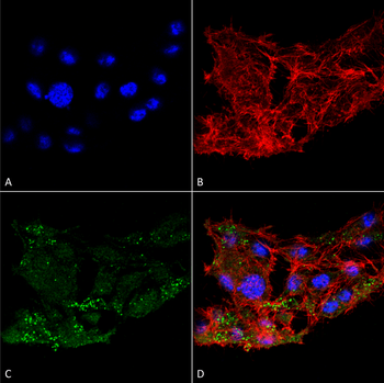
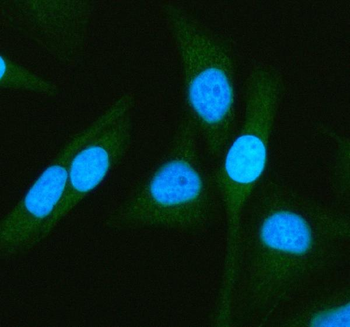
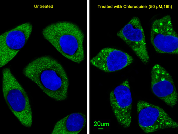
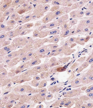
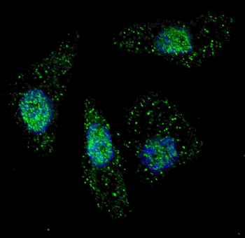
Immunocytochemistry/Immunofluorescence analysis using Rabbit Anti-ATG7 Polyclonal Antibody. Tissue: Colon carcinoma cell line (RKO). Species: Human. Fixation: 4% Formaldehyde for 15 min at RT. Primary Antibody: Rabbit Anti-ATG7 Polyclonal Antibody at 1:100 for 60 min at RT. Secondary Antibody: Goat Anti-Rabbit ATTO 488 at 1:100 for 60 min at RT. Counterstain: Phalloidin Texas Red F-Actin stain; DAPI (blue) nuclear stain at 1:1000, 1:5000 for 60 min at RT, 5 min at RT. Localization: Cytoplasm, Preautophagosomal Structure, Organelle membrane. Magnification: 60X. (A) DAPI nuclear stain. (B) Phalloidin Texas Red F-Actin stain. (C) ATG7 Antibody. (D) Composite.

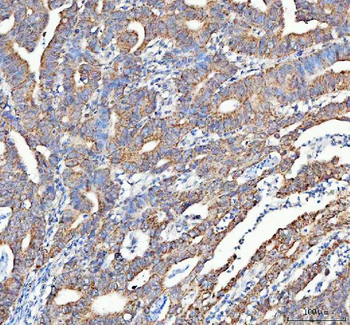
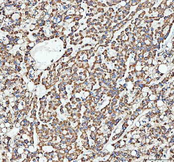
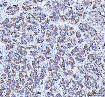
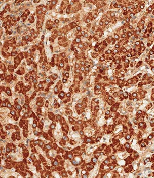
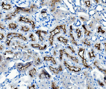
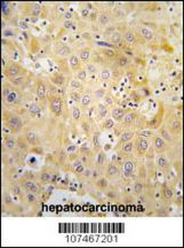
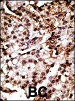
Immunohistochemistry analysis using Rabbit Anti-ATG10 Polyclonal Antibody. Tissue: Brain. Species: Human. Fixation: Formalin Fixed Paraffin-Embedded. Primary Antibody: Rabbit Anti-ATG10 Polyclonal Antibody at 1:50 for 31 min at RT. Counterstain: Hematoxylin. Magnification: 10X.

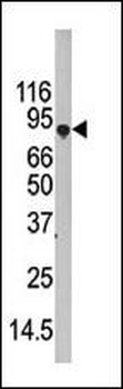
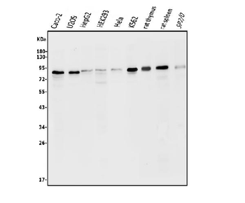
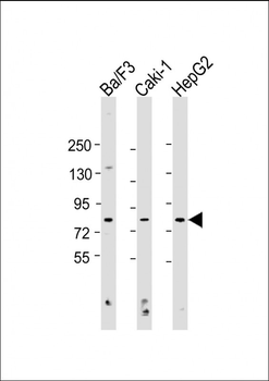
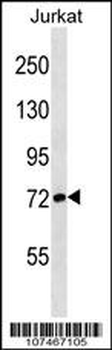
Western blot analysis of Rat brain cell lysates showing detection of ~77.9 kDa ATG7 protein using Rabbit Anti-ATG7 Polyclonal Antibody. Lane 1: Molecular Weight Ladder (MW). Lane 2: Rat brain cell lysates. Load: 20 μg. Block: 2% BSA and 2% Skim Milk in 1X TBST. Primary Antibody: Rabbit Anti-ATG7 Polyclonal Antibody at 1:1000 for 16 hours at 4°C. Secondary Antibody: Goat Anti-Rabbit IgG: HRP at 1:2000 for 60 min at RT. Color Development: ECL solution for 6 min at RT. Predicted/Observed Size: ~77.9 kDa.

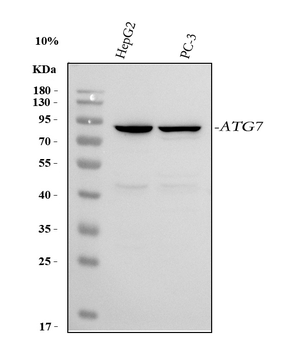
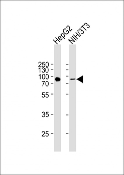
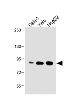
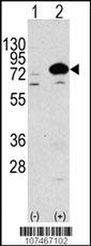
Western blot analysis of Human Cervical cancer cell line (HeLa) lysate showing detection of ~77.9 kDa ATG7 protein using Rabbit Anti-ATG7 Polyclonal Antibody. Lane 1: Molecular Weight Ladder (MW). Lane 2: Human Cervical cancer cell line (HeLa) lysate. Load: 15 μg. Block: 5% Skim Milk in 1X TBST. Primary Antibody: Rabbit Anti-ATG7 Polyclonal Antibody at 1:1000 for 60 min at RT. Secondary Antibody: Goat Anti-Rabbit IgG: HRP at 1:2000 for 60 min at RT. Color Development: ECL solution for 6 min at RT. Predicted/Observed Size: ~77.9 kDa.
Quick Database Links
UniProt Details
− No UniProt data available
NCBI Gene Details
− No NCBI Gene data available
NCBI Reference Sequences
−Associated Accession Numbers
Curated reference sequences for the gene transcript and protein product| Protein | NP_001129503.2 |
|---|
Documents Download
Datasheet
Product Information
Request a Document
Protocol Information
WB
Western Blot (IB, immunoblot)
IHC
Immunohistochemistry
IF
Immunofluorescence
ICC
Immunocytochemistry
ATG7 Antibody (orb54993)

- 0.0
Based on 0 reviews
Participating in our Biorbyt product reviews program enables you to support fellow scientists by sharing your firsthand experience with our products.
Login to Submit a ReviewAvailable Sizes
Select a size below