You have no items in your shopping cart.
Description
Research Area
Cell Biology
Images & Validation
−Item 1 of 3
| Tested Applications | ICC, IF, WB |
|---|---|
| Dilution Range | WB (1:1000); ICC/IF (1:100) |
| Reactivity | Human, Mouse |
| Application Notes |
Key Properties
−| Host | Rabbit |
|---|---|
| Clonality | Polyclonal |
| Immunogen | Synthetic peptide from the C-terminal of human ATG2A |
| Target | ATG2A |
| Molecular Weight | 90-100kDa |
| Purification | Peptide Affinity Purified |
| Conjugation | Biotin |
Storage & Handling
−| Storage | Conjugated antibodies should be stored according to the product label |
|---|---|
| Buffer/Preservatives | 136.36mM Ethanolamine, 133.23 mM Chlorides, 9.55mM Phosphates, 9.55mM Sodium Bicarbonate. |
| Concentration | 1 mg/ml |
| Expiration Date | 12 months from date of receipt. |
| Disclaimer | For research use only |
Alternative Names
−ATG2A, KIAA0404, ATG2 autophagy related homolog A, ATG2a_HUMAN, BC023754, 1810013C15Rik
Similar Products
−
ATG2A Rabbit Polyclonal Antibody (Biotin) [orb2105452]
WB
Bovine, Canine, Equine, Guinea pig, Human, Mouse, Porcine, Rabbit, Rat, Zebrafish
Rabbit
Polyclonal
Biotin
100 μl
Quality Guarantee
Explore bioreagents carefree to elevate your research. All our products are rigorously tested for performance. If a product does not perform as described on its datasheet, our scientific support team will provide expert troubleshooting, a prompt replacement, or a refund. For full details, please see our Terms & Conditions and Buying Guide. Contact us at [email protected].
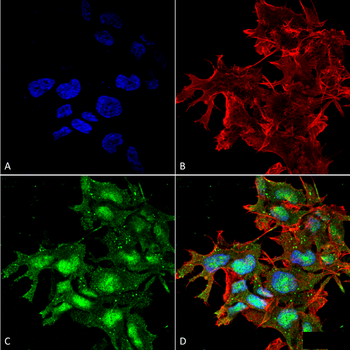
Immunocytochemistry/Immunofluorescence analysis using Rabbit Anti-ATG2A Polyclonal Antibody. Tissue: Neuroblastoma cell line (SK-N-BE). Species: Human. Fixation: 4% Formaldehyde for 15 min at RT. Primary Antibody: Rabbit Anti-ATG2A Polyclonal Antibody at 1:100 for 60 min at RT. Secondary Antibody: Goat Anti-Rabbit ATTO 488 at 1:100 for 60 min at RT. Counterstain: Phalloidin Texas Red F-Actin stain; DAPI (blue) nuclear stain at 1:1000, 1:5000 for 60min RT, 5min RT. Localization: Nucleus, Cytoplasm. Magnification: 60X. (A) DAPI (blue) nuclear stain (B) Phalloidin Texas Red F-Actin stain (C) ATG2A Antibody (D) Composite.
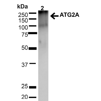
Western blot analysis of Mouse Brain showing detection of ~212.9kDa ATG2A protein using Rabbit Anti-ATG2A Polyclonal Antibody. Lane 1: MW Ladder. Lane 2: Mouse Brain (20 μg). Load: 20 μg. Block: 5% milk + TBST for 1 hour at RT. Primary Antibody: Rabbit Anti-ATG2A Polyclonal Antibody at 1:1000 for 1 hour at RT. Secondary Antibody: Goat Anti-Rabbit: HRP at 1:2000 for 1 hour at RT. Color Development: TMB solution for 12 min at RT. Predicted/Observed Size: ~212.9kDa.
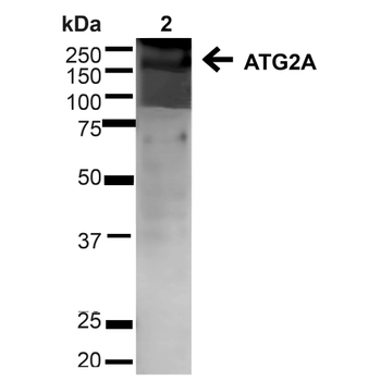
Western blot analysis of Human Cervical cancer cell line (HeLa) lysate showing detection of ~212.9kDa ATG2A protein using Rabbit Anti-ATG2A Polyclonal Antibody. Lane 1: MW Ladder. Lane 2: Human HeLa (20 μg). Load: 20 μg. Block: 5% milk + TBST for 1 hour at RT. Primary Antibody: Rabbit Anti-ATG2A Polyclonal Antibody at 1:1000 for 1 hour at RT. Secondary Antibody: Goat Anti-Rabbit: HRP at 1:2000 for 1 hour at RT. Color Development: TMB solution for 12 min at RT. Predicted/Observed Size: ~212.9kDa.
Quick Database Links
UniProt Details
− No UniProt data available
NCBI Gene Details
− No NCBI Gene data available
NCBI Reference Sequences
−Associated Accession Numbers
Curated reference sequences for the gene transcript and protein product| Protein | NP_055919.2 |
|---|
Documents Download
Datasheet
Product Information
Request a Document
Protocol Information
WB
Western Blot (IB, immunoblot)
IF
Immunofluorescence
ICC
Immunocytochemistry
ATG2A Antibody (Biotin) (orb377369)

- 0.0
Based on 0 reviews
Participating in our Biorbyt product reviews program enables you to support fellow scientists by sharing your firsthand experience with our products.
Login to Submit a ReviewAvailable Sizes
Select a size below
Choose Conjugation or Carrier Free Version
Free Secondary Antibody (20 ul)0/0
Please add an antibody product to your cart first.