You have no items in your shopping cart.
APOLIPOPROTEIN B Antibody
SKU: orb345277
Description
Images & Validation
−Item 1 of 1
| Tested Applications | ELISA, IHC, IP, WB |
|---|---|
| Dilution Range | ELISA: 1:2,000 - 1:10,000, IHC: 1:50 - 1:500, IP: 1:100, WB: 1:200 - 1:1,000 |
| Reactivity | Human |
| Application Notes |
Key Properties
−| Antibody Type | Primary Antibody |
|---|---|
| Host | Goat |
| Clonality | Polyclonal |
| Isotype | IgG |
| Immunogen | apoLipoprotein Type B was isolated from human plasma by density gradient centrifugation followed by HPLC purification. |
| Purity | This product has been prepared by immunoaffinity chromatography using immobilized antigens followed by extensive cross-adsorption against other apoLipoproteins and human serum proteins to remove any unwanted specificities. Typically less than 1% cross reactivity against other types of apoLipoprotein was detected by ELISA against purified standards. This antibody reacts with human apoLipoprotein B and has negligible cross-reactivity with Type A-I, A-II, C-I, C-II, C-III, E and J apoLipoproteins. Specific cross reaction of anti-apoLipoprotein antibodies with antigens from other species has not been determined. Non-specific cross reaction of anti-apoLipoprotein antibodies with other human serum proteins is negligible. |
| Conjugation | Unconjugated |
Storage & Handling
−| Storage | Store vial at 4° C prior to opening. This product is stable 4° C as an undiluted liquid. Dilute only prior to immediate use. For extended storage mix with an equal volume of glycerol, aliquot contents and freeze at -20° C or below. Avoid cycles of freezing and thawing. |
|---|---|
| Form/Appearance | Liquid (sterile filtered) |
| Buffer/Preservatives | Preservative: 0.01% (w/v) Sodium Azide. Stabilizer: None; Buffer: 0.125 M Sodium Borate, 0.075 M Sodium Chloride, 0.005 M EDTA, pH 8.0 |
| Concentration | 1.0 mg/mL |
| Expiration Date | 12 months from date of receipt. |
| Disclaimer | For research use only |
Alternative Names
−goat anti-Apolipoprotein B antibody, Apo B-100, Apo B-48, APOB protein antibody, Apolipoprotein-B 100 antibody, Apolipoprotein B 48 antibody, Apolipoprotein B antibody, FLDB antibody
Similar Products
−- Item 1 of 4
- Item 1 of 5
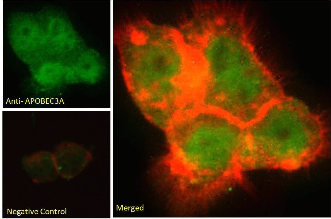
Goat anti-Phorbolin 1 / APOBEC3A Antibody [orb19763]
ELISA, FC, IF, IHC, WB
Human
Goat
Polyclonal
Unconjugated
100 μg - Item 1 of 1
- Item 1 of 3
APOB Antibody [orb1410492]
IHC
Human
Mouse
Monoclonal
Unconjugated
20 μg, 100 μg, 100 μg (without BSA and Azide) - Item 1 of 3
APOB Antibody [orb1410439]
IHC
Human
Mouse
Monoclonal
Unconjugated
20 μg, 100 μg, 100 μg (without BSA and Azide)


Quality Guarantee
Explore bioreagents carefree to elevate your research. All our products are rigorously tested for performance. If a product does not perform as described on its datasheet, our scientific support team will provide expert troubleshooting, a prompt replacement, or a refund. For full details, please see our Terms & Conditions and Buying Guide. Contact us at [email protected].
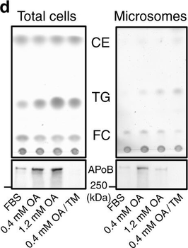
LDs are present in the lumen of the type I NR. a Huh7 treated with OA/TM for 24–48 h harbored LDs in the lumen of the ER (left; arrowheads) and within the type I NR (right; arrows mark the NR). INM: inner nuclear membrane, ONM: outer nuclear membrane. Bars, 0.2 µm. b Huh7-expressing HRP-KDEL treated with OA/TM for 24 h. DAB precipitated in the type I NR lumen (arrows). Bar, 0.5 µm. c Mouse hepatocytes in vivo after high-fat diet feeding for 6 weeks and TM injection. Lumenal LDs were observed in the ER (arrowheads), the nuclear cistern (arrowhead in the inset) (left figure; Bar, 0.5 µm), and in the type I NR (arrows mark the NR) (right figure; Bar, 0.2 µm). They contained more nuclear LDs than the control fed the high-fat diet and injected with vehicle alone. Mean ± SD of three independent experiments. *p < 0.01, Student's t test. d Microsomes of Huh7 treated with none, 0.4 mm OA, 1.2 mm OA, or OA/TM for 48 h. The OA/TM-treated cell microsome contained triglycerides (TG) and cholesterol esters (CE) most abundantly (by thin layer chromatography), but showed the lowest amount of ApoB (by Western blotting). e Three different kinds of LDs in the nuclear area: Nucleoplasmic LDs (A), NR-lumenal LDs (within the type I NR) (B), and cytoplasmic LDs (within the type II NR) (C). f Nucleoplasmic LDs (arrowheads) and NR-lumenal LDs (arrows) are distinguished by whether they are outside of or within LBR rings, respectively. Huh7 treated with OA/TM for 48 h. Both LDs were reduced by MTPi (100 nm BAY 13-9952). Bar, 10 µm. See also Supplementary Fig. 2e. g The number of nucleoplasmic LDs, NR-lumenal LDs, and cytoplasmic LDs within the type II NR were counted in randomly taken electron micrographs of Huh7 treated with OA/TM for 48–72 h. Box plot of pooled data from three independent experiments. The average is shown by +. Number of nuclei examined = 128.
Quick Database Links
UniProt
RefSeq:AAH51278.1
UniProt Details
− No UniProt data available
NCBI Reference Sequences
−Associated Accession Numbers
Curated reference sequences for the gene transcript and protein product| RefSeq | AAH51278.1 |
|---|
Documents Download
Datasheet
Product Information
Request a Document
Protocol Information
WB
Western Blot (IB, immunoblot)
IHC
Immunohistochemistry
ELISA
Enzyme-linked Immunosorbent Assay (EIA)
IP
Immunoprecipitation
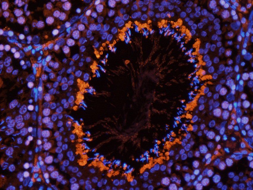
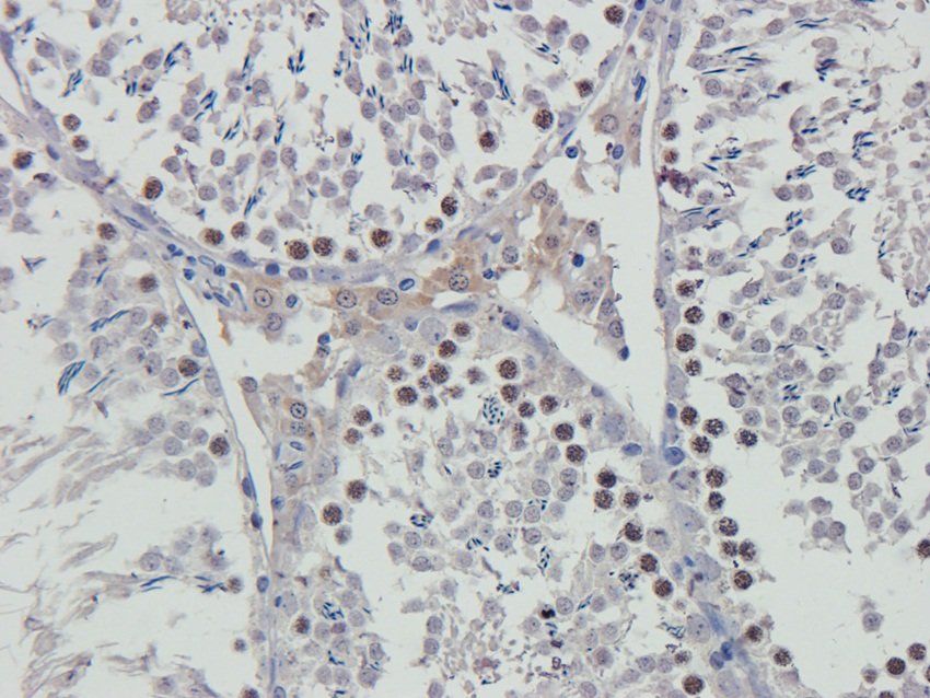
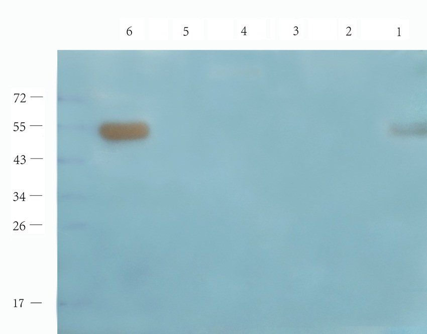
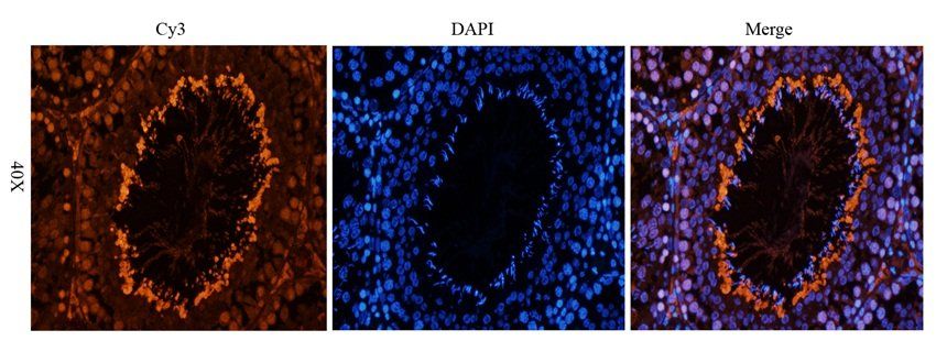
APOLIPOPROTEIN B Antibody (orb345277)

- 0.0
Based on 0 reviews
Participating in our Biorbyt product reviews program enables you to support fellow scientists by sharing your firsthand experience with our products.
Login to Submit a ReviewAvailable Sizes
Select a size below