You have no items in your shopping cart.
Description
Research Area
Metabolism Research
Images & Validation
−
Item 1 of 10
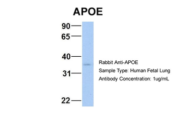
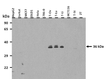
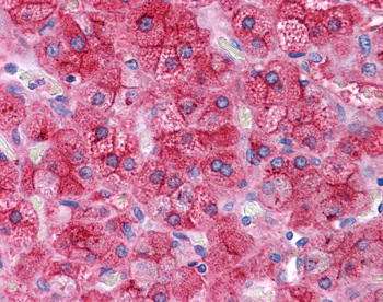
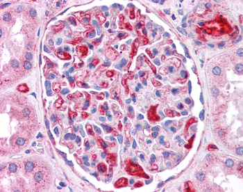
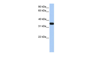
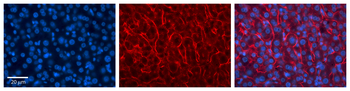
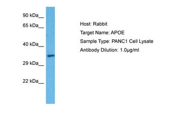
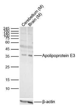
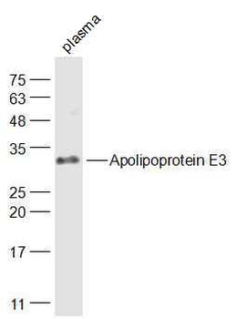
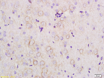
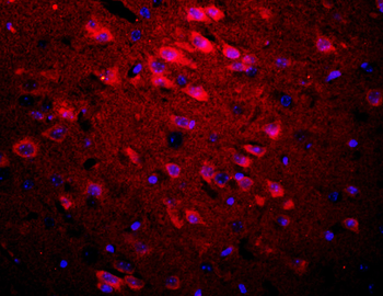
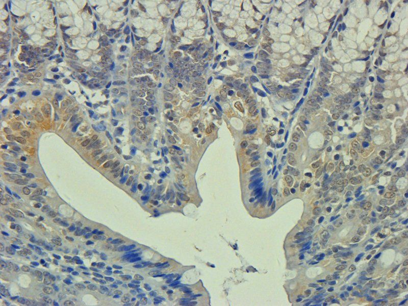
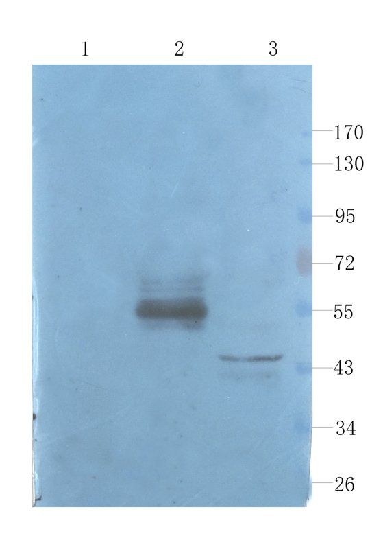
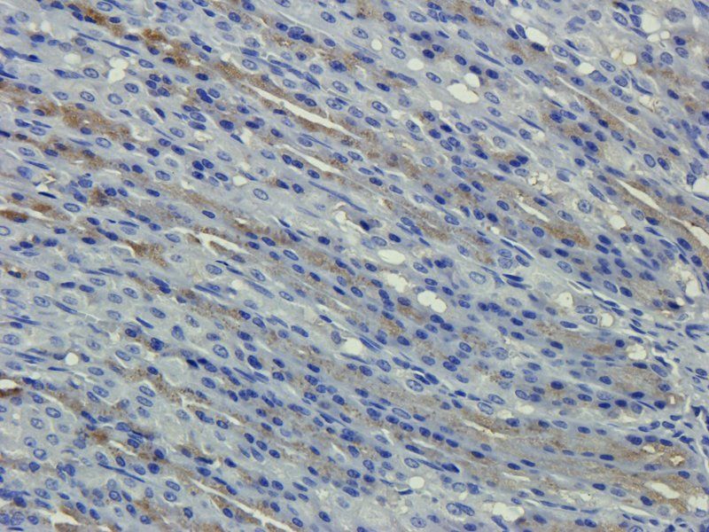
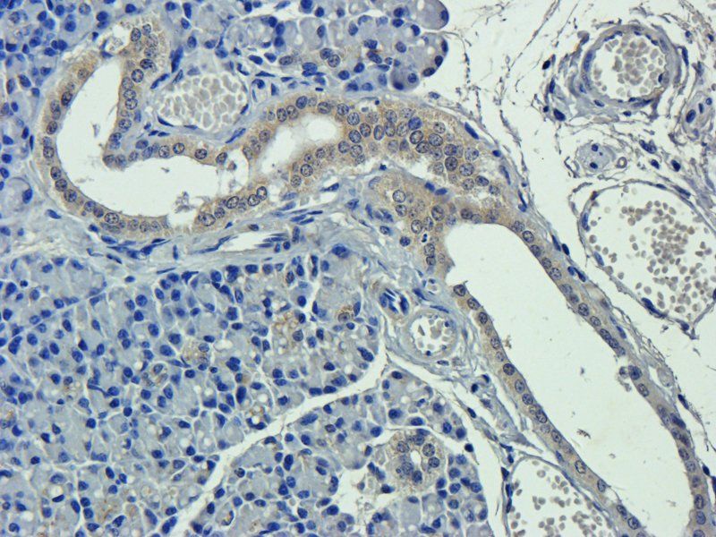
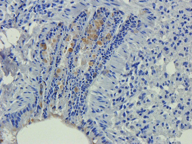
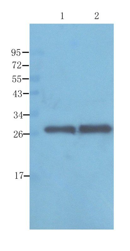
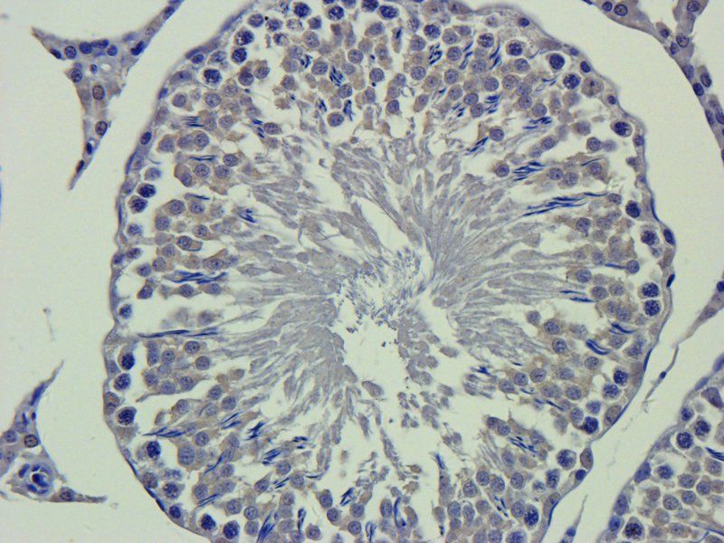
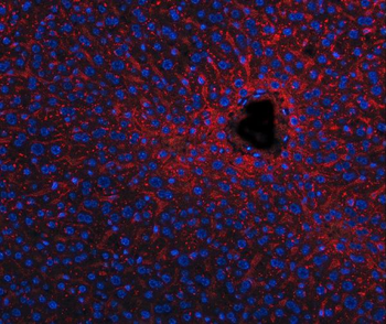
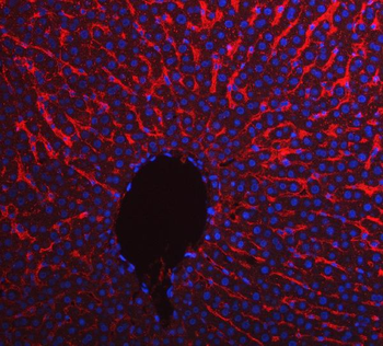
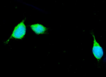
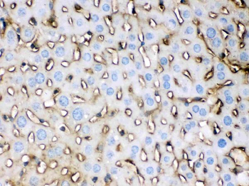
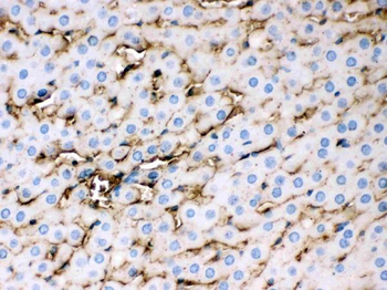
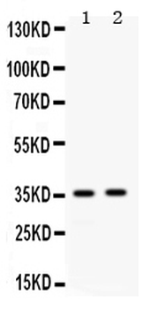
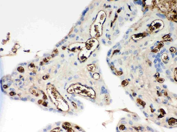
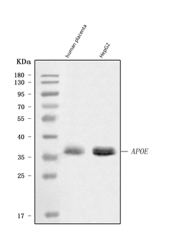
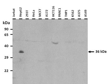
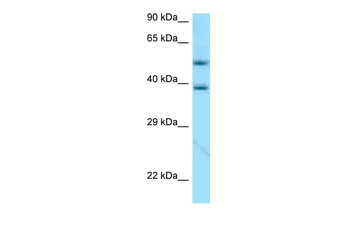
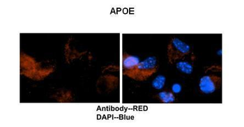
| Tested Applications | IHC, WB |
|---|---|
| Reactivity | Human, Mouse, Rat |
| Predicted Reactivity | Human |
Key Properties
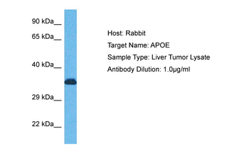
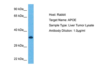
−| Host | Rabbit |
|---|---|
| Clonality | Polyclonal |
| Immunogen | The immunogen is a synthetic peptide directed towards the N terminal region of human APOE |
| Target | APOE |
| Protein Sequence | Synthetic peptide located within the following region: KVLWAALLVTFLAGCQAKVEQAVETEPEPELRQQTEWQSGQRWELALGRF |
| Molecular Weight | 36 kDa |
| Purification | Affinity Purified |
| Conjugation | Unconjugated |
Storage & Handling
−| Storage | Maintain refrigerated at 2-8°C for up to 2 weeks. For long term storage store at -20°C in small aliquots to prevent freeze-thaw cycles. |
|---|---|
| Buffer/Preservatives | Liquid. Purified antibody supplied in 1x PBS buffer with 0.09% (w/v) sodium azide and 2% sucrose. |
| Concentration | 0.5 mg/ml |
| Expiration Date | 12 months from date of receipt. |
| Disclaimer | For research use only |
Alternative Names
−Anti-AD2 antibody, Anti-Apo-E antibody, Anti-APOE antibody, Anti-APOE_HUMAN antibody, Anti-APOEA antibody, Anti-Apolipoprotein E antibody, Anti-Apolipoprotein E3 antibody, Anti-ApolipoproteinE antibody, Anti-Apoprotein antibody, Anti-LDLCQ5 antibody, Anti-LPG antibody
Similar Products
−- Item 1 of 6
Apolipoprotein E3 Rabbit Polyclonal Antibody [orb4523]
IF, IHC-Fr, IHC-P, WB
Bovine, Porcine
Human, Mouse, Rat
Rabbit
Polyclonal
Unconjugated
50 μl, 100 μl, 200 μl - Item 1 of 7
Apolipoprotein E Rabbit Polyclonal Antibody [orb339614]
IHC-P, WB
Human, Rat
Rabbit
Polyclonal
Unconjugated
100 μg, 500 μg - Item 1 of 6
Apolipoprotein E/APOE Rabbit Polyclonal Antibody [orb371720]
ICC, IF, IHC, WB
Mouse, Rat
Rabbit
Polyclonal
Unconjugated
100 μg - Item 1 of 5
Apolipoprotein E/APOE Rabbit Polyclonal Antibody [orb251510]
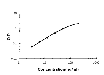
ELISA, FC, IF, IHC, WB
Human
Rabbit
Polyclonal
Unconjugated
100 μg - Item 1 of 4
APOE Rabbit Polyclonal Antibody [orb333723]
IHC, WB
Rat, Zebrafish
Human, Mouse
Rabbit
Polyclonal
Unconjugated
100 μl


Quality Guarantee
Explore bioreagents carefree to elevate your research. All our products are rigorously tested for performance. If a product does not perform as described on its datasheet, our scientific support team will provide expert troubleshooting, a prompt replacement, or a refund. For full details, please see our Terms & Conditions and Buying Guide. Contact us at [email protected].
Protocol Information
WB
Western Blot (IB, immunoblot)
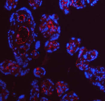
IHC
Immunohistochemistry
Available Sizes
Select a size below
Free Secondary Antibody (20 ul)0/0
Please add an antibody product to your cart first.