You have no items in your shopping cart.
Description
Images & Validation
−Item 1 of 2
| Tested Applications | ELISA, IHC, WB |
|---|---|
| Dilution Range | ELISA: 1:2,000 - 1:10,000, IHC: 5.0 µg/ml, WB: 1:200 - 1:1,000 |
| Reactivity | Human |
| Application Notes |
Key Properties
−| Antibody Type | Primary Antibody |
|---|---|
| Host | Rabbit |
| Clonality | Polyclonal |
| Isotype | IgG |
| Immunogen | This affinity purified antibody was prepared from whole rabbit serum produced by repeated immunizations with a synthetic peptide corresponding to an internal region surrounding pS377 of Human Apc1 protein. |
| Target | ANAPC1 |
| Purity | This product is an affinity purified antibody produced by immunoaffinity chromatography using phospho peptide coupled to agarose beads followed by solid phase adsorption(s) against non-phospho peptide and non-specific peptide to remove any unwanted reactivities. This antibody is specific for phosphorylated human APC1 protein at the pS377 residue. A BLAST analysis was used to suggest reactivity with this protein from human, dog, rat, and bovine based on 100% homology for the immunogen sequence. Cross reactivity with APC1 protein from mouse is expected, as this sequence show 90% homologous between human and mouse. Cross reactivity with APC1 homologues from other sources has not been determined. Minimal reactivity is expected with the non-phosphorylated form of the protein. |
| Conjugation | Unconjugated |
Storage & Handling
−| Storage | Store vial at -20° C prior to opening. Aliquot contents and freeze at -20° C or below for extended storage. Avoid cycles of freezing and thawing. Centrifuge product if not completely clear after standing at room temperature. This product is stable for several weeks at 4° C as an undiluted liquid. Dilute only prior to immediate use. |
|---|---|
| Form/Appearance | Liquid (sterile filtered) |
| Buffer/Preservatives | Preservative: 0.01% (w/v) Sodium Azide. Stabilizer: None; Buffer: 0.02 M Potassium Phosphate, 0.15 M Sodium Chloride, pH 7.2 |
| Concentration | 0.21 mg/mL |
| Expiration Date | 12 months from date of receipt. |
| Dry Ice Shipping | Please note: This product requires shipment on dry ice. A dry ice surcharge will apply. |
| Disclaimer | For research use only |
Alternative Names
−rabbit anti-APC1 pS377 Antibody, APC-1, APC 1, ANAPC1 antibody, Anaphase promoting complex subunit 1 antibody, Cyclosome subunit 1 antibody, MCPR antibody, Meiotic checkpoint regulator antibody
Similar Products
−- Item 1 of 4
APC1/ANAPC1 Rabbit Polyclonal Antibody [orb1939945]
ELISA, FC, ICC, IF, IHC, WB
Human
Rabbit
Polyclonal
Unconjugated
100 μg - Item 1 of 2
APC1 rabbit pAb Antibody [orb770068]
ELISA, IF, IHC
Human, Mouse, Rat
Polyclonal
Unconjugated
50 μl, 100 μl - Item 1 of 3
ANAPC1 (Phospho-S688) Rabbit Polyclonal Antibody [orb382450]
IF, IHC, WB
Human, Mouse
Rabbit
Polyclonal
Unconjugated
30 μl, 100 μl, 200 μl, 50 μl - Item 1 of 2
Phospho-APC1 (Ser355) Rabbit Polyclonal Antibody [orb4393]
IF, IHC-Fr, IHC-P, WB
Bovine, Canine, Equine, Human, Porcine, Rabbit, Sheep
Mouse, Rat
Rabbit
Polyclonal
Unconjugated
50 μl, 100 μl, 200 μl - Item 1 of 2
ANAPC1 Rabbit Polyclonal Antibody [orb394721]
ELISA, IHC, WB
Human, Mouse, Rat
Rabbit
Polyclonal
Unconjugated
50 μg, 100 μg


Quality Guarantee
Explore bioreagents carefree to elevate your research. All our products are rigorously tested for performance. If a product does not perform as described on its datasheet, our scientific support team will provide expert troubleshooting, a prompt replacement, or a refund. For full details, please see our Terms & Conditions and Buying Guide. Contact us at [email protected].
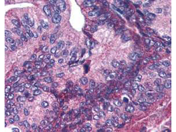
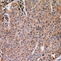
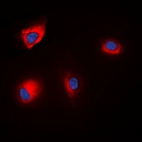
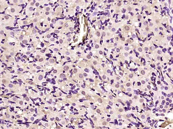
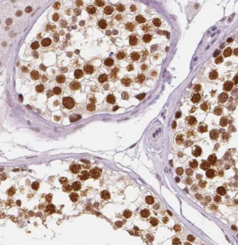
Biorbyt's affinity purified anti-APC1 pS377 antibody was used at 5.0 µg/ml to detect signal in a variety of tissues including multi-human, multi-brain and multi-cancer slides. This image shows moderate positive cytoplasmic and occasional nuclear staining of pancreatic carcinoma cells at 60X. Tissue was formalin-fixed and paraffin embedded. The image shows localization of the antibody as the precipitated red signal, with a hematoxylin purple nuclear counterstain.
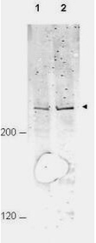
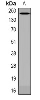
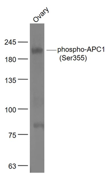
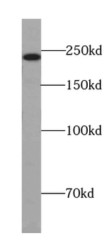
Western blot using Biorbyt's Affinity Purified anti-APC1 pS377 antibody shows detection of a band ~215 kDa corresponding to phosphorylated human APC1 (arrowhead). Lane 1 shows lysate from asynchronous cells. Lane 2 shows lysate from cells treated with nocodazole. While some phosphorylated APC1 is present in untreated cell, the amount of phosphorylated protein is increased in cell preparations arrested in mitosis. Each lane contains approximately 30 ug of HeLa whole cell lysates, separated by 4-8% SDS-PAGE followed by transfer to nitrocellulose. After blocking the membrane was probed with the primary antibody diluted to 1:1000 overnight at 4°C followed by washes and reaction with a 1:10000 dilution of IRDye800 conjugated Gt-a-Rabbit IgG [H&L] MX for 45 min at room temperature.
Documents Download
Datasheet
Product Information
Request a Document
Protocol Information
WB
Western Blot (IB, immunoblot)
IHC
Immunohistochemistry
ELISA
Enzyme-linked Immunosorbent Assay (EIA)
ANAPC1 Antibody (orb345507)

- 0.0
Based on 0 reviews
Participating in our Biorbyt product reviews program enables you to support fellow scientists by sharing your firsthand experience with our products.
Login to Submit a ReviewAvailable Sizes
Select a size below
Choose Conjugation or Carrier Free Version
Free Secondary Antibody (20 ul)0/0
Please add an antibody product to your cart first.