You have no items in your shopping cart.
On Promotion
Species Host
Tested Applications
Reactivity
Conjugation
Unconjugated 180FITC 17Biotin 16HRP 14APC 11PE 8PerCP 8AP 5Cy3 5APC/Cy5.5 4APC/Cy7 4BF350 4BF405 4BF488 4BF555 4BF594 4BF647 4BF680 4BF700 4BF750 4Cy5 4Cy5.5 4Cy7 4PE/Cy5 4PE/Cy5.5 4PE/Cy7 4Pacific Blue 4PerCP/Cy5.5 4PerCP/Cy7 4RBITC 4RPE 4AE 3IRDye800 3Fluoro488 2Fluoro550 2Fluoro594 2Fluoro647 2SureLight 488 2iFluor488 2iFluor568 1
Featured Product
Search results for: 'single domain'
- Featured
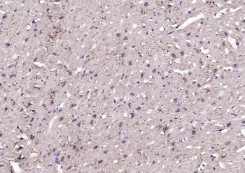
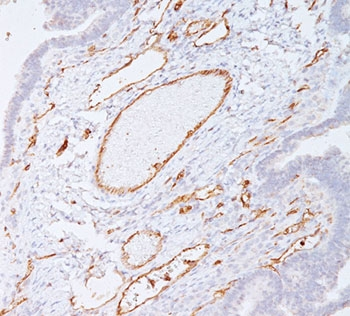
 Item 1 of 16DBPA Rabbit Polyclonal Antibody [orb156545]Featured
IF, IHC-Fr, IHC-P, WB
Human, Mouse, Rat
Bovine, Canine, Porcine, Rabbit, Sheep
Rabbit
Polyclonal
Unconjugated
200 μl, 100 μl, 50 μl - Item 1 of 9
- Item 1 of 9
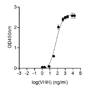
- Item 1 of 4GFP and other derivatives of GFP Antibody [orb1562805]
ELISA, IP, WB
Jellyfish
Camelus
Monoclonal
Unconjugated
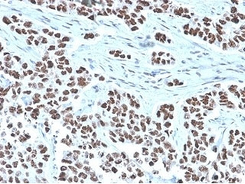
50 μg, 1000 μg, 2000 μg, 100 μg, 250 μg - Item 1 of 6NFIA Antibody / Nuclear Factor 1 A [orb2634961]
FACS, IF, IHC-P, WB
Human
Mouse
Monoclonal
Unconjugated
100 μg - Item 1 of 6NFIA Antibody / Nuclear Factor 1 A [orb2634962]
FACS, IF, IHC-P, WB
Human
Mouse
Monoclonal
Unconjugated
100 μg, 20 μg - Item 1 of 6
- Item 1 of 4
ELISA, IF, IHC, WB
Human
Camelus
Monoclonal
Unconjugated
100 μg, 250 μg, 1000 μg, 2000 μg, 50 μg - Item 1 of 6
ELISA, NeA
Virus
Camelus
Recombinant
Unconjugated
0.1 mg, 0.02 mg - Item 1 of 6


![SARS-CoV-2 (COVID-19) Spike Neutralization Single Domain Antibody [E10]](/images/pub/media/catalog/product/NewWebsite/15/orb1274179_1.jpg)
