You have no items in your shopping cart.
Phospho-DNA PKcs PRKDC/DNA Rabbit Monoclonal Antibody
SKU: orb548611
Description
Research Area
Epigenetics & Chromatin, Molecular Biology
Images & Validation
−Item 1 of 3
| Tested Applications | ICC, IF, IHC, WB |
|---|---|
| Dilution Range | WB 1:500-2000 IHC 1:50-200 ICC/IF 1:50-200 |
| Reactivity | Human |
Key Properties
−| Antibody Type | Primary Antibody |
|---|---|
| Host | Rabbit |
| Clonality | Monoclonal |
| Isotype | Rabbit IgG |
| Clone No. | BDQFX38 |
| Immunogen | A synthesized peptide derived from human Phospho-DNA PKcs (S2056) |
| Target | DNA-dependent protein kinase catalytic subunit |
| Molecular Weight | 185 kDa |
| Purification | Affinity-chromatography |
| Conjugation | Unconjugated |
Storage & Handling
−| Storage | Maintain refrigerated at 2-8°C for up to 2 weeks. For long term storage store at -20°C in small aliquots to prevent freeze-thaw cycles. |
|---|---|
| Form/Appearance | Liquid |
| Buffer/Preservatives | Rabbit IgG in stabilizing components, phosphate buffered saline, pH 7.4, 150mM NaCl, 0.02% sodium azide and 50% glycerol. *This antibody is supplied in a stabilized formulation. Compatibility with conjugation reactions depends on the chemistry of the conjugation method used. For conjugation methods that are not compatible with the stabilizing components present in this formulation, a carrier-free antibody format is required. |
| Concentration | 0.5mg/ml |
| Expiration Date | 12 months from date of receipt. |
| Disclaimer | For research use only |
Alternative Names
−DNA-dependent protein kinase catalytic subunit; DNA-PK catalytic subunit; DNA-PKcs; 2.7.11.1; DNPK1; p460; PRKDC; HYRC, HYRC1
Quality Guarantee
Explore bioreagents carefree to elevate your research. All our products are rigorously tested for performance. If a product does not perform as described on its datasheet, our scientific support team will provide expert troubleshooting, a prompt replacement, or a refund. For full details, please see our Terms & Conditions and Buying Guide. Contact us at [email protected].
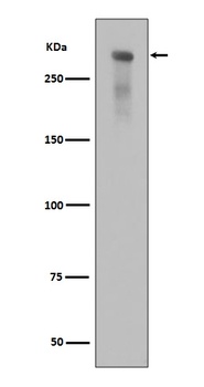
Western blot analysis of Phospho-DNA PKcs (Ser2056) expression in alkaline treated Jurkat cell lysate.
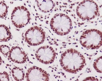
Immunohistochemical analysis of paraffin-embedded human colon tissue using anti-Phospho-DNA PKcs (S2056) antibody. The section was pre-treated using heat mediated antigen retrieval with Tris-EDTA buffer (pH 9.0) for 20 minutes.The tissues were blocked in 5% BSA for 30 minutes at room temperature, washed with ddH2O and PBS, and then probed with the primary antibody (1/200) for 30 minutes at room temperature. The detection was performed using an HRP conjugated compact polymer system. DAB was used as the chromogen. Tissues were counterstained with hematoxylin and mounted with DPX.
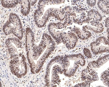
Immunohistochemical analysis of paraffin-embedded human colon carcinoma tissue using anti-Phospho-DNA PKcs (S2056) antibody. The section was pre-treated using heat mediated antigen retrieval with Tris-EDTA buffer (pH 9.0) for 20 minutes.The tissues were blocked in 5% BSA for 30 minutes at room temperature, washed with ddH2O and PBS, and then probed with the primary antibody (1/200) for 30 minutes at room temperature. The detection was performed using an HRP conjugated compact polymer system. DAB was used as the chromogen. Tissues were counterstained with hematoxylin and mounted with DPX.
Quick Database Links
Gene Symbol
DNA-dependent protein kinase catalytic subunit
UniProt
UniProt Details
− No UniProt data available
Documents Download
Datasheet
Product Information
Request a Document
Protocol Information
WB
Western Blot (IB, immunoblot)
IHC
Immunohistochemistry
IF
Immunofluorescence
ICC
Immunocytochemistry
Phospho-DNA PKcs PRKDC/DNA Rabbit Monoclonal Antibody (orb548611)

- 0.0
Based on 0 reviews
Participating in our Biorbyt product reviews program enables you to support fellow scientists by sharing your firsthand experience with our products.
Login to Submit a ReviewAvailable Sizes
Select a size below
Free Secondary Antibody (20 ul)0/0
Please add an antibody product to your cart first.