You have no items in your shopping cart.
KO/KD
Validated
Validated
Description
Research Area
Cell Biology, Metabolism Research, Signal Transduction
Images & Validation
−
Item 1 of 6
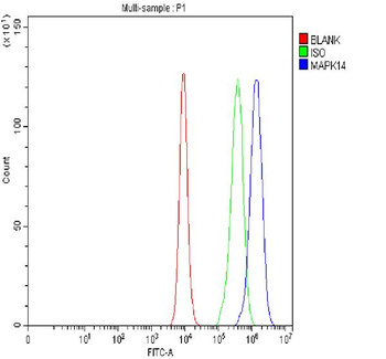
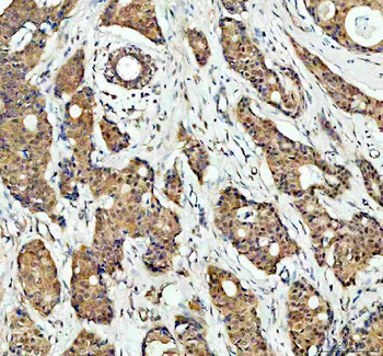
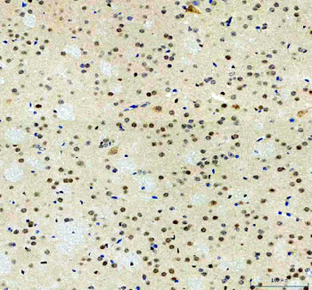
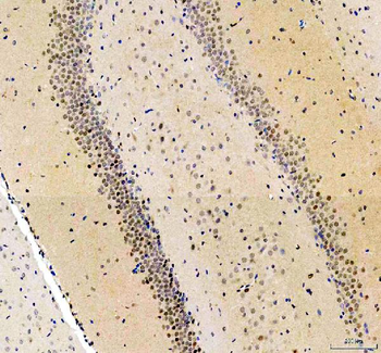
| Tested Applications | FC, ICC, IHC, IP, KO/KD Validated, WB |
|---|---|
| Dilution Range | Western blot, 0.25-0.5 μg/ml, Human, Mouse, Rat Immunohistochemistry (Paraffin-embedded Section), 2-5 μg/ml, Human, Mouse, Rat Immunocytochemistry/Immunofluorescence, 5 μg/ml, Human Immunoprecipitation, 0.5-2 μg/ml, Human Flow Cytometry (Fixed), 1-3 μg/1x10^6 cells, Human |
| Reactivity | Human, Mouse, Rat |
Related Conjugates & Formulations
−Key Properties
−| Antibody Type | Primary Antibody |
|---|---|
| Host | Rabbit |
| Clonality | Polyclonal |
| Isotype | Rabbit IgG |
| Immunogen | A synthetic peptide corresponding to a sequence at the C-terminus of human p38 alpha/MAPK14, identical to the related mouse and rat sequences. |
| Target | Mitogen-activated protein kinase 14 |
| Molecular Weight | 38 kDa |
| Purification | Immunogen affinity purified. |
| Conjugation | Unconjugated |
Storage & Handling
−| Storage | Maintain refrigerated at 2-8°C for up to 2 weeks. For long term storage store at -20°C in small aliquots to prevent freeze-thaw cycles. |
|---|---|
| Form/Appearance | Lyophilized |
| Buffer/Preservatives | Each vial contains 4 mg Trehalose, 0.9 mg NaCl, 0.2 mg Na2HPO4. |
| Concentration | Adding 0.2 ml of distilled water will yield a concentration of 500 μg/ml. |
| Expiration Date | 12 months from date of receipt. |
| Disclaimer | For research use only |
Alternative Names
−Mannose-6-phosphate isomerase; 5.3.1.8; Phosphohexomutase; Phosphomannose isomerase; PMI; MPI; PMI1
Similar Products
−
p38 alpha/MAPK14 Rabbit Polyclonal Antibody (Fluoro647) [orb3119679]
Human, Mouse, Rat
Rabbit
Polyclonal
Fluoro647
100 μgp38 alpha/MAPK14 Rabbit Polyclonal Antibody (Fluoro550) [orb3119681]
Human, Mouse, Rat
Rabbit
Polyclonal
Fluoro550
100 μgp38 alpha/MAPK14 Rabbit Polyclonal Antibody (Fluoro488) [orb3119682]
Human, Mouse, Rat
Rabbit
Polyclonal
Fluoro488
100 μgp38 alpha/MAPK14 Rabbit Polyclonal Antibody (Fluoro594) [orb3119680]
Human, Mouse, Rat
Rabbit
Polyclonal
Fluoro594
100 μgp38 alpha/MAPK14 Rabbit Polyclonal Antibody (FITC) [orb2597262]
Human, Mouse, Rat
Rabbit
Polyclonal
FITC
100 μg
Quality Guarantee
Explore bioreagents carefree to elevate your research. All our products are rigorously tested for performance. If a product does not perform as described on its datasheet, our scientific support team will provide expert troubleshooting, a prompt replacement, or a refund. For full details, please see our Terms & Conditions and Buying Guide. Contact us at [email protected].
Quick Database Links
Gene Symbol
Mitogen-activated protein kinase 14
UniProt
UniProt Details
− No UniProt data available
Protocol Information
WB
Western Blot (IB, immunoblot)
IHC
Immunohistochemistry
FC
Flow Cytometry
ICC
Immunocytochemistry
IP
Immunoprecipitation
Available Sizes
Select a size below
Free Secondary Antibody (20 ul)0/0
Please add an antibody product to your cart first.