You have no items in your shopping cart.
Myeloperoxidase/MPO Rabbit Polyclonal Antibody (APC)
SKU: orb2580005
Description
Research Area
Cancer Biology, Cardiovascular Research, Immunology & Inflammation, Metabolism Research
Images & Validation
−Item 1 of 4
| Tested Applications | FC |
|---|---|
| Dilution Range | Flow Cytometry, Optimal dilutions should be determined by end users. |
| Reactivity | Human, Mouse, Rat |
Related Conjugates & Formulations
−Key Properties
−| Antibody Type | Primary Antibody |
|---|---|
| Host | Rabbit |
| Clonality | Polyclonal |
| Isotype | Rabbit IgG |
| Immunogen | A synthetic peptide corresponding to a sequence at the C-terminus of human MPO, different from the related mouse and rat sequences by one amino acid. |
| Target | Myeloperoxidase |
| Molecular Weight | 83869 Da |
| Purification | Immunogen affinity purified. |
| Conjugation | APC |
Storage & Handling
−| Storage | At -20°C for one year from date of receipt. Avoid repeated freezing and thawing. Protect from light. |
|---|---|
| Form/Appearance | Liquid |
| Buffer/Preservatives | Each vial contains 50% glycerol, 0.9% NaCl, 0.2% Na2HPO4, 0.02% NaN3. |
| Expiration Date | 12 months from date of receipt. |
| Disclaimer | For research use only |
Alternative Names
−Myeloperoxidase; MPO; 1.11.2.2; Myeloperoxidase; 89 kDa myeloperoxidase; 84 kDa myeloperoxidase; Myeloperoxidase light chain; Myeloperoxidase heavy chain; MPO
Similar Products
−
MPO Rabbit Polyclonal Antibody (APC) [orb992096]
ICC, IF
Canine, Equine, Guinea pig, Rabbit, Rat
Human, Mouse
Rabbit
Polyclonal
APC
100 μlMPO Rabbit Polyclonal Antibody (APC-Cy7) [orb1606766]
ICC, IF
Canine, Equine, Guinea pig, Rabbit, Rat
Human, Mouse
Rabbit
Polyclonal
APC/Cy7
100 μlMPO Rabbit Polyclonal Antibody (APC-Cy5.5) [orb1606767]
ICC, IF
Canine, Equine, Guinea pig, Rabbit, Rat
Human, Mouse
Rabbit
Polyclonal
APC/Cy5.5
100 μl
Quality Guarantee
Explore bioreagents carefree to elevate your research. All our products are rigorously tested for performance. If a product does not perform as described on its datasheet, our scientific support team will provide expert troubleshooting, a prompt replacement, or a refund. For full details, please see our Terms & Conditions and Buying Guide. Contact us at [email protected].
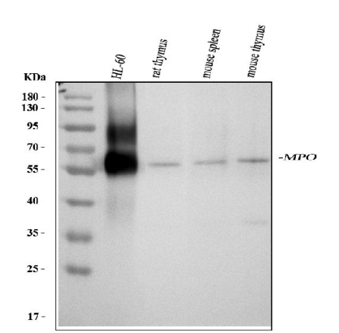
Western blot analysis of MPO using anti-MPO antibody. Electrophoresis was performed on a 5-20% SDS-PAGE gel at 70V (Stacking gel) / 90V (Resolving gel) for 2-3 hours. The sample well of each lane was loaded with 30 ug of sample under reducing conditions
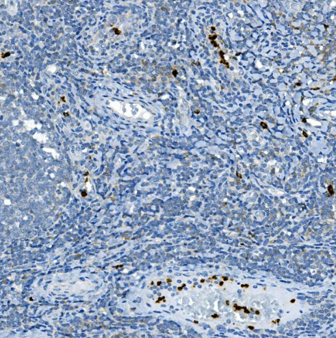
IHC analysis of MPO using anti-MPO antibody. MPO was detected in a paraffin-embedded section of human tonsil tissue. Heat mediated antigen retrieval was performed in EDTA buffer (pH 8.0, epitope retrieval solution). The tissue section was blocked with 10% goat serum. The tissue section was then incubated with 2 μg/ml rabbit anti-MPO Antibody overnight at 4°C. Peroxidase Conjugated Goat Anti-rabbit IgG was used as secondary antibody and incubated for 30 minutes at 37°C. The tissue section was developed using HRP Conjugated Rabbit IgG Super Vision Assay Kit with DAB as the chromogen
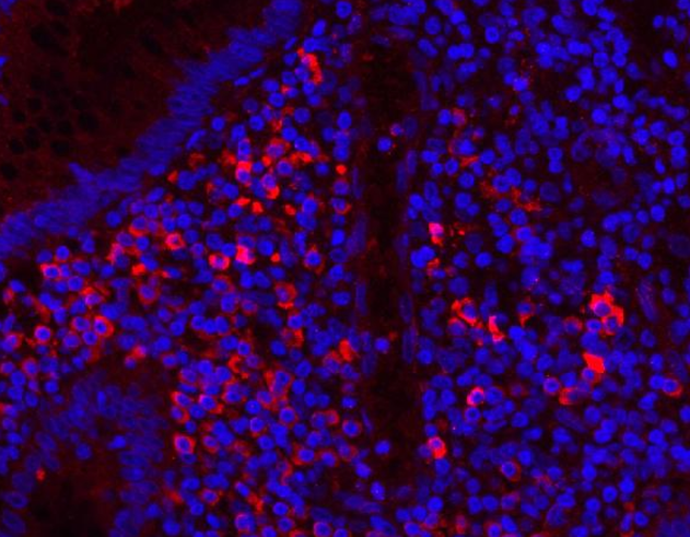
MPO was detected in a paraffin-embedded section of human appendicitis tissue. Heat mediated antigen retrieval was performed in EDTA buffer (pH 8.0, epitope retrieval solution). The tissue section was blocked with 10% goat serum. The tissue section was then incubated with 5 μg/mL rabbit anti-MPO Antibody overnight at 4°C. Cy3 Conjugated Goat Anti-Rabbit IgG was used as secondary antibody at 1:500 dilution and incubated for 30 minutes at 37°C. The section was counterstained with DAPI. Visualize using a fluorescence microscope and filter sets appropriate for the label used
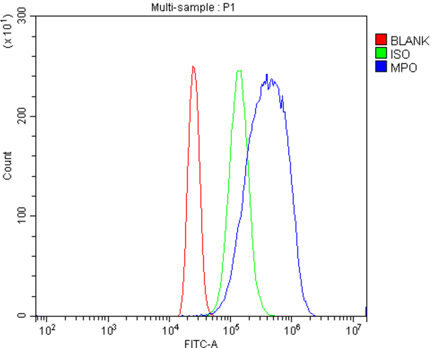
Flow Cytometry analysis of HL-60 cells using anti-MPO antibody. Overlay histogram showing HL-60 cells stained with orb2580005 (Blue line). To facilitate intracellular staining, cells were fixed with 4% paraformaldehyde and permeabilized with permeabilization buffer. The cells were blocked with 10% normal goat serum. And then incubated with rabbit anti-MPO Antibody for 30 min at 20°C. DyLight®488 conjugated goat anti-rabbit IgG was used as secondary antibody for 30 minutes at 20°C. Isotype control antibody (Green line) was rabbit IgG (1 μg/1x106) used under the same conditions. Unlabelled sample without incubation with primary antibody and secondary antibody (Red line) was used as a blank control
Quick Database Links
Gene Symbol
Myeloperoxidase
UniProt
UniProt Details
− No UniProt data available
Documents Download
Datasheet
Product Information
Request a Document
Myeloperoxidase/MPO Rabbit Polyclonal Antibody (APC) (orb2580005)

- 0.0
Based on 0 reviews
Participating in our Biorbyt product reviews program enables you to support fellow scientists by sharing your firsthand experience with our products.
Login to Submit a ReviewAvailable Sizes
Select a size below
Free Secondary Antibody (20 ul)0/0
Please add an antibody product to your cart first.