You have no items in your shopping cart.
mGLUR6 Antibody
SKU: orb2651424
Description
Research Area
Cell Biology
Images & Validation
−Item 1 of 2
| Tested Applications | IF, WB |
|---|---|
| Dilution Range | WB: WB (1/500 - 1/1000), IF/IC (1/100 - 1/500), IF: WB (1/500 - 1/1000), IF/IC (1/100 - 1/500) |
| Reactivity | Human, Mouse, Rat |
Key Properties
−| Host | Rabbit |
|---|---|
| Clonality | Polyclonal |
| Clone No. | GRM6 |
| Conjugation | Unconjugated |
Storage & Handling
−| Storage | Maintain refrigerated at 2-8°C for up to 2 weeks. For long term storage store at -20°C in small aliquots to prevent freeze-thaw cycles. |
|---|---|
| Expiration Date | 12 months from date of receipt. |
| Disclaimer | For research use only |
Alternative Names
−GPRC1F; MGLUR6; Metabotropic glutamate receptor 6; mGluR6
Similar Products
−- Item 1 of 4
GRM6 Rabbit Polyclonal Antibody [orb574284]
WB
Bovine, Canine, Equine, Guinea pig, Rabbit, Rat, Yeast, Zebrafish
Human, Mouse
Rabbit
Polyclonal
Unconjugated
100 μl - Item 1 of 2
Metabotropic Glutamate Receptor 6 (GPRC1F) Antibody (C-term) [orb1934887]
FC, WB
Human
Rabbit
Polyclonal
Unconjugated
400 μl - Item 1 of 3
- Item 1 of 2
mGLUR6 Rabbit Polyclonal Antibody [orb214014]
IF, WB
Human, Mouse, Rat
Rabbit
Polyclonal
Unconjugated
30 μl, 100 μl, 200 μl, 50 μl - Item 1 of 1


Quality Guarantee
Explore bioreagents carefree to elevate your research. All our products are rigorously tested for performance. If a product does not perform as described on its datasheet, our scientific support team will provide expert troubleshooting, a prompt replacement, or a refund. For full details, please see our Terms & Conditions and Buying Guide. Contact us at [email protected].

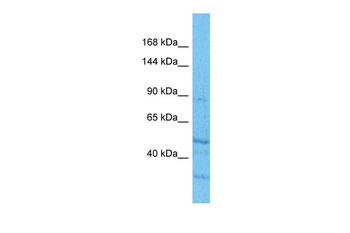
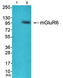
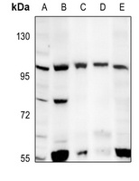
Western blot analysis of mGLUR6 expression in C6 (A), SP20 (B), SHSY5Y (C), U87MG (D), A375 (E) whole cell lysates.
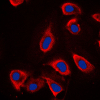
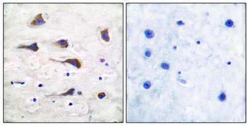
Immunofluorescent analysis of mGLUR6 staining in NIH3T3 cells. Formalin-fixed cells were permeabilized with 0.1% Triton X-100 in TBS for 5-10 minutes and blocked with 3% BSA-PBS for 30 minutes at room temperature. Cells were probed with the primary antibody in 3% BSA-PBS and incubated overnight at 4°C in a humidified chamber. Cells were washed with PBST and incubated with a DyLight 594-conjugated secondary antibody (red) in PBS at room temperature in the dark. DAPI was used to stain the cell nuclei (blue).
Quick Database Links
UniProt
UniProt Details
− No UniProt data available
Documents Download
Datasheet
Product Information
Request a Document
Protocol Information
WB
Western Blot (IB, immunoblot)
IF
Immunofluorescence
mGLUR6 Antibody (orb2651424)

- 0.0
Based on 0 reviews
Participating in our Biorbyt product reviews program enables you to support fellow scientists by sharing your firsthand experience with our products.
Login to Submit a ReviewAvailable Sizes
Select a size below