You have no items in your shopping cart.
Description
Research Area
Cell Biology, Disease Biomarkers, Immunology & Inflammation
Images & Validation
−Item 1 of 2
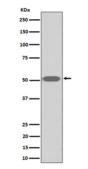
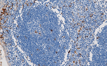
| Tested Applications | ICC, IF, IHC, IP, WB |
|---|---|
| Dilution Range | WB 1:1000-5000 IHC 1:50-200 ICC/IF 1:50-200 IP 1:20 |
| Reactivity | Human, Mouse, Rat |
Key Properties
−| Antibody Type | Primary Antibody |
|---|---|
| Host | Rabbit |
| Clonality | Monoclonal |
| Isotype | Rabbit IgG |
| Clone No. | BGDQX34 |
| Immunogen | A synthesized peptide derived from human Lysozyme |
| Target | Lysozyme C |
| Molecular Weight | 15 kDa |
| Purification | Affinity-chromatography |
| Conjugation | Unconjugated |
Storage & Handling
−| Storage | Maintain refrigerated at 2-8°C for up to 2 weeks. For long term storage store at -20°C in small aliquots to prevent freeze-thaw cycles. |
|---|---|
| Form/Appearance | Liquid |
| Buffer/Preservatives | Rabbit IgG in stabilizing components, phosphate buffered saline, pH 7.4, 150mM NaCl, 0.02% sodium azide and 50% glycerol. *This antibody is supplied in a stabilized formulation. Compatibility with conjugation reactions depends on the chemistry of the conjugation method used. For conjugation methods that are not compatible with the stabilizing components present in this formulation, a carrier-free antibody format is required. |
| Concentration | 0.5mg/ml |
| Expiration Date | 12 months from date of receipt. |
| Disclaimer | For research use only |
Alternative Names
−Lysozyme C; 3.2.1.17; 1, 4-beta-N-acetylmuramidase C; LYZ; LZM
Similar Products
−- Item 1 of 4
Lysozyme Recombinant Rabbit Monoclonal Antibody [orb1499338]
IF, IHC-Fr, IHC-P, WB
Human, Mouse
Rabbit
Recombinant
Unconjugated
50 μl, 100 μl, 25 μl - Item 1 of 1
Thromboxane A2 receptor Rabbit Monoclonal Antibody [orb866831]
WB
Human, Mouse, Rat
Rabbit
Monoclonal
Unconjugated
100 μl - Item 1 of 1
Quality Guarantee
Explore bioreagents carefree to elevate your research. All our products are rigorously tested for performance. If a product does not perform as described on its datasheet, our scientific support team will provide expert troubleshooting, a prompt replacement, or a refund. For full details, please see our Terms & Conditions and Buying Guide. Contact us at [email protected].
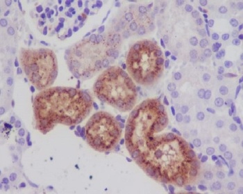
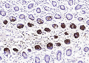
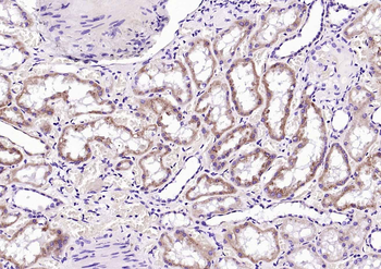
Immunohistochemical analysis of paraffin-embedded mouse kidney, using Lysozyme Antibody.
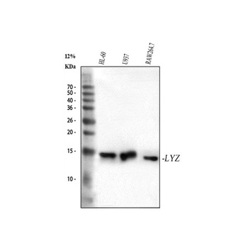
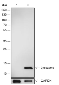
Western blot analysis of Lysozyme using anti-Lysozyme antibody (orb547865). Electrophoresis was performed on a 5-20% SDS-PAGE gel at 70V (Stacking gel)/90V (Resolving gel) for 2-3 hours. The sample well of each lane was loaded with 30 ug of sample under reducing conditions. Lane 1: human HL-60 whole cell lysates, Lane 2: human U937 whole cell lysates, Lane 3: mouse RAW264.7 whole cell lysates. After electrophoresis, proteins were transferred to a nitrocellulose membrane at 150 mA for 50-90 minutes. Blocked the membrane with 5% non-fat milk/TBS for 1.5 hour at RT. The membrane was incubated with mouse anti-Lysozyme antigen affinity purified monoclonal antibody (Catalog # orb547865) at 1:5000 overnight at 4°C, then washed with TBS-0.1%Tween 3 times with 5 minutes each and probed with a goat anti-mouse IgG-HRP secondary antibody at a dilution of 1:5000 for 1.5 hour at RT. The signal is developed using an Enhanced Chemiluminescent detection (ECL) kit (Catalog # orb90502) with Tanon 5200 system. A specific band was detected for Lysozyme at approximately 15 kDa. The expected band size for Lysozyme is at 17 kDa.
Quick Database Links
Gene Symbol
Lysozyme C
UniProt
UniProt Details
− No UniProt data available
Documents Download
Datasheet
Product Information
Request a Document
Protocol Information
WB
Western Blot (IB, immunoblot)
IHC
Immunohistochemistry
IF
Immunofluorescence
ICC
Immunocytochemistry
IP
Immunoprecipitation
Lysozyme LYZ Rabbit Monoclonal Antibody (orb547865)

- 0.0
Based on 0 reviews
Participating in our Biorbyt product reviews program enables you to support fellow scientists by sharing your firsthand experience with our products.
Login to Submit a ReviewAvailable Sizes
Select a size below
Free Secondary Antibody (20 ul)0/0
Please add an antibody product to your cart first.