You have no items in your shopping cart.
Description
Research Area
Epigenetics, Immunology
Images & Validation
−Item 1 of 4
| Tested Applications | FC, IP, WB |
|---|---|
| Reactivity | Human |
| Application Notes |
Key Properties
−| Antibody Type | Primary Antibody |
|---|---|
| Clonality | Monoclonal |
| Isotype | Mouse IgG1 |
| Clone No. | LAT-01 |
| Immunogen | Bacterially produced recombinant polypeptide corresponding to the entire cytoplasmic domain of human LAT. |
| Target | LAT |
| Purification | Purified by protein-A affinity chromatography. |
| Conjugation | Unconjugated |
Storage & Handling
−| Storage | Maintain refrigerated at 2-8°C for up to 2 weeks. For long term storage store at -20°C in small aliquots to prevent freeze-thaw cycles. |
|---|---|
| Buffer/Preservatives | Phosphate buffered saline (PBS), pH 7.4, 15 mM sodium azide |
| Concentration | 1 mg/ml |
| Expiration Date | 12 months from date of receipt. |
| Disclaimer | For research use only |
Alternative Names
−IMD52
Similar Products
−- Item 1 of 10
Lat Rabbit Polyclonal Antibody [orb1939864]
ELISA, FC, IHC, WB
Mouse, Rat
Rabbit
Polyclonal
Unconjugated
100 μg - Item 1 of 1
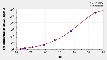
Human Linker for Activation of T-Cell (LAT) ELISA Kit [orb775956]
Human
0.32-20 ng/mL
0.105 ng/mL
96 T, 48 T - Item 1 of 3
LAT rabbit pAb Antibody [orb768410]
ELISA, IHC, WB
Human, Mouse, Rat
Polyclonal
Unconjugated
50 μl, 100 μl - Item 1 of 3
LAT rabbit pAb Antibody [orb765577]
ELISA, IHC, WB
Human, Mouse, Rat
Polyclonal
Unconjugated
50 μl, 100 μl - Item 1 of 3


Quality Guarantee
Explore bioreagents carefree to elevate your research. All our products are rigorously tested for performance. If a product does not perform as described on its datasheet, our scientific support team will provide expert troubleshooting, a prompt replacement, or a refund. For full details, please see our Terms & Conditions and Buying Guide. Contact us at [email protected].

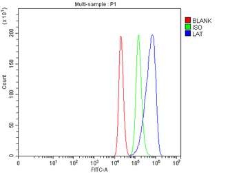
Flow cytometry multicolor intracellular staining of human peripheral whole blood stained using anti-LAT (LAT-01) purified antibody (concentration in sample 1 µg/ml, GAM APC) and anti-human CD3 (UCHT1) Pacific Blue™ antibody (20 µl reagent / 100 µl of peripheral whole blood).
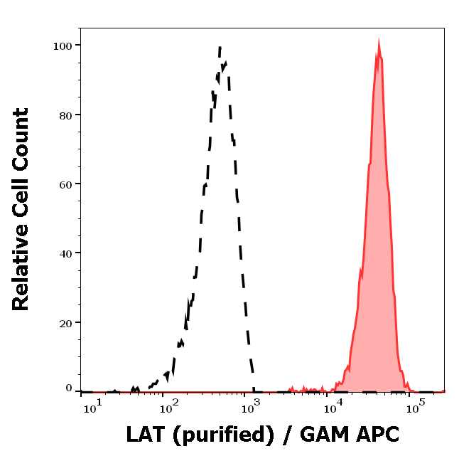
Separation of human CD3 positive LAT positive lymphocytes (red-filled) from neutrophil granulocytes (black-dashed) in flow cytometry analysis (intracellular staining) of peripheral whole blood stained using anti-LAT (LAT-01) purified antibody (concentration in sample 1 µg/ml, GAM APC).
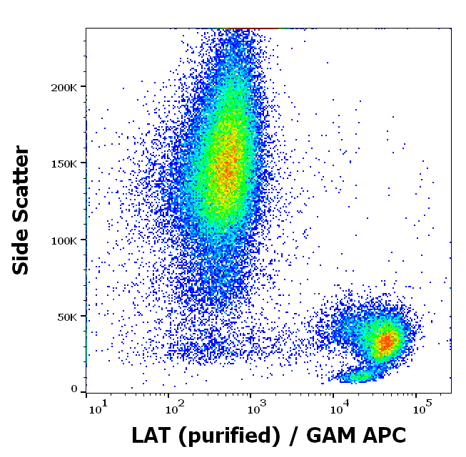
Flow cytometry intracellular staining pattern of human peripheral whole blood using anti-LAT (LAT-01) purified antibody (concentration in sample 1 µg/ml, GAM APC).
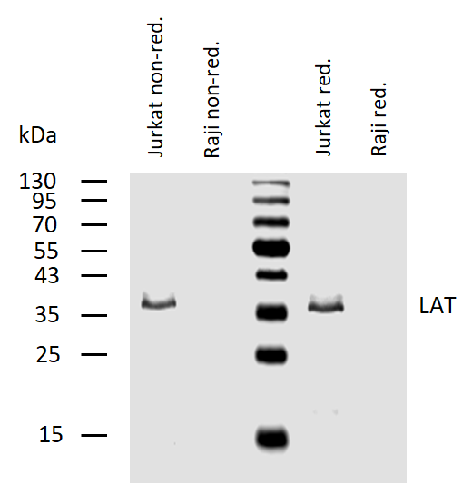
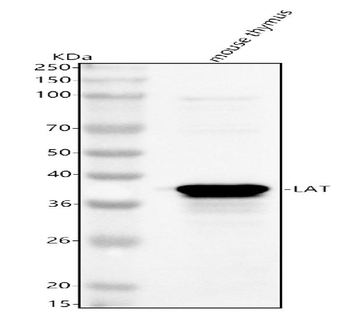
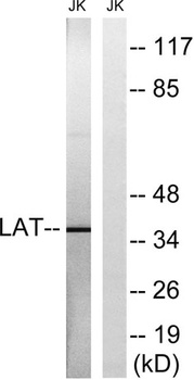
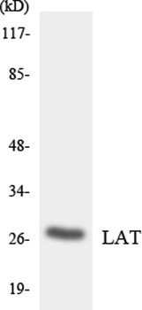
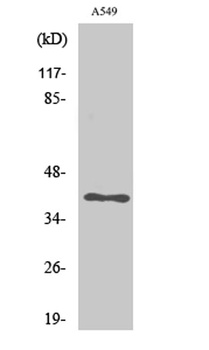
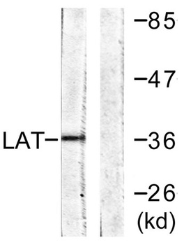
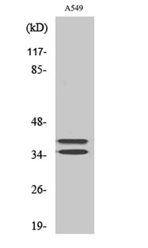
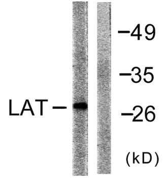
Western blotting analysis of human LAT using mouse monoclonal antibody LAT-01 in lysates of Jurkat cells (positive) and Raji cells (negative control) under non-reducing and reducing conditions. Nitrocellulose membrane was probed with 2 µg/ml of mouse anti-LAT monoclonal antibody followed by IRDye800-conjugated anti-mouse secondary antibody. A specific band was detected for LAT at approximately 38 kDa.
Documents Download
Datasheet
Product Information
Request a Document
Protocol Information
WB
Western Blot (IB, immunoblot)
FC
Flow Cytometry
IP
Immunoprecipitation
LAT Antibody (orb43896)

- 0.0
Based on 0 reviews
Participating in our Biorbyt product reviews program enables you to support fellow scientists by sharing your firsthand experience with our products.
Login to Submit a ReviewAvailable Sizes
Select a size below
Free Secondary Antibody (20 ul)0/0
Please add an antibody product to your cart first.