You have no items in your shopping cart.
Description
Research Area
Immunology & Inflammation
Images & Validation
−Item 1 of 3
| Tested Applications | FC, IHC-P, IP, WB |
|---|---|
| Reactivity | Human |
| Application Notes |
Key Properties
−| Antibody Type | Primary Antibody |
|---|---|
| Clonality | Monoclonal |
| Isotype | Mouse IgG1 |
| Clone No. | MEM-98 |
| Immunogen | Human CD6 antigen purified by immunoaffinity chromatography from HBP-ALL cells followed by preparative SDS-PAGE of non-boiled non-reduced sample (excised piece of gel corresponding to the 100 kDa zone). |
| Target | CD6 |
| Purification | Purified by protein-A affinity chromatography. |
| Conjugation | Unconjugated |
Storage & Handling
−| Storage | Maintain refrigerated at 2-8°C for up to 2 weeks. For long term storage store at -20°C in small aliquots to prevent freeze-thaw cycles. |
|---|---|
| Buffer/Preservatives | Phosphate buffered saline (PBS), pH 7.4, 15 mM sodium azide |
| Concentration | 1 mg/ml |
| Expiration Date | 12 months from date of receipt. |
| Disclaimer | For research use only |
Alternative Names
−T12, TP120
Similar Products
−- Item 1 of 7
- Item 1 of 6
- Item 1 of 3
CD6 Antibody [orb1410566]
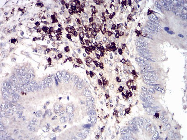
IHC
Human
Rabbit
Monoclonal
Unconjugated
20 μg, 100 μg, 100 μg (without BSA and Azide) 


Quality Guarantee
Explore bioreagents carefree to elevate your research. All our products are rigorously tested for performance. If a product does not perform as described on its datasheet, our scientific support team will provide expert troubleshooting, a prompt replacement, or a refund. For full details, please see our Terms & Conditions and Buying Guide. Contact us at [email protected].

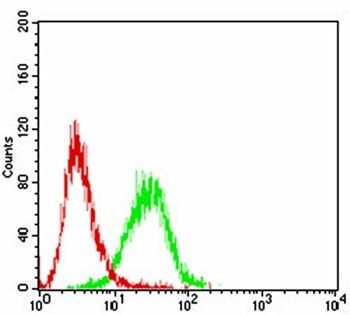
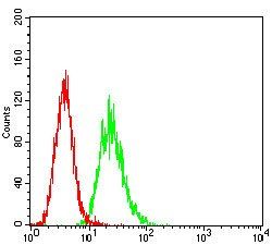
Separation of human CD6 positive lymphocytes (red-filled) from neutrophil granulocytes (black-dashed) in flow cytometry analysis (surface staining) of peripheral whole blood stained using anti-human CD6 (MEM-98) purified antibody (concentration in sample 2 μg/ml, GAM APC).
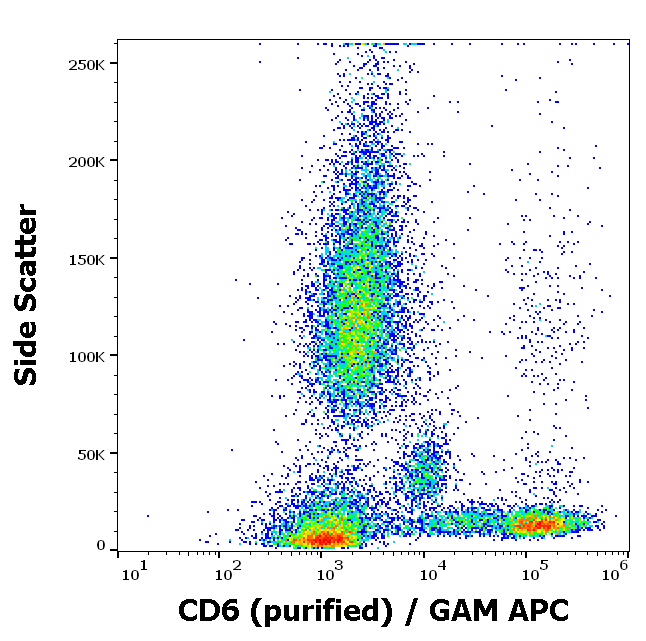
Flow cytometry surface staining pattern of human peripheral whole blood stained using anti-human CD6 (MEM-98) purified antibody (concentration in sample 2 μg/ml, GAM APC).
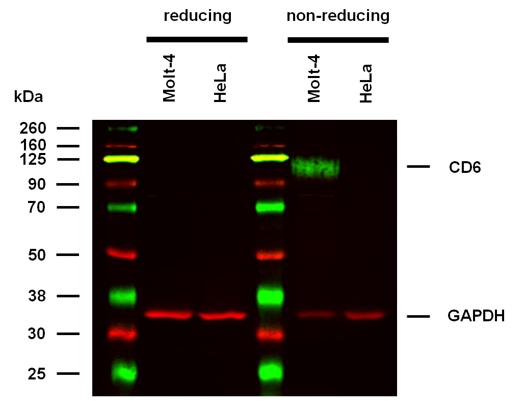
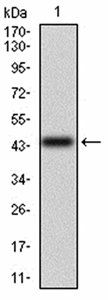
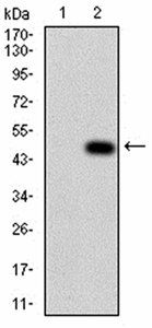
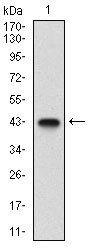
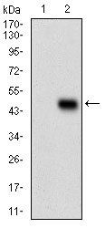
Anti-Hu CD6 Purified (clone MEM-98) works in WB application under non-reducing conditions. Western blotting analysis was performed on whole cell extracts (RIPA lysis buffer) of Molt-4 and HeLa cell lines, mixed and heated (100°C, 5 min) with reducing and non-reducing SDS-loading buffer. Samples were resolved using 10% Tris-glycine SDS gel electrophoresis. Nitrocellulose membrane blot was probed with mouse IgG1 monoclonal antibody MEM-98 (1 µg/ml), followed by IRDye 800CW Goat-anti-Mouse IgG (green). Mouse anti-GAPDH monoclonal antibody FF26A conjugated with DyLight 680 (0.1 µg/ml), was used as the loading control (red). Multiplex fluorescent Western blot detection was performed. CD6 molecules were detected at ~100 kDa in Molt-4 cell line under non-reducing conditions.
Documents Download
Datasheet
Product Information
Request a Document
Protocol Information
WB
Western Blot (IB, immunoblot)
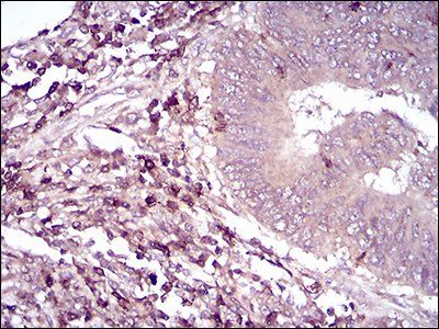
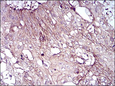
IHC-P
Immunohistochemistry Paraffin
FC
Flow Cytometry
IP
Immunoprecipitation
CD6 Antibody (orb44614)

- 0.0
Based on 0 reviews
Participating in our Biorbyt product reviews program enables you to support fellow scientists by sharing your firsthand experience with our products.
Login to Submit a ReviewAvailable Sizes
Select a size below
Free Secondary Antibody (20 ul)0/0
Please add an antibody product to your cart first.