You have no items in your shopping cart.
Description
Research Area
Immunology & Inflammation
Images & Validation
−Item 1 of 4
| Tested Applications | FC, ICC, IHC, IP, WB |
|---|---|
| Reactivity | Human |
| Application Notes |
Key Properties
−| Antibody Type | Primary Antibody |
|---|---|
| Clonality | Monoclonal |
| Isotype | Mouse IgG2a kappa |
| Clone No. | L17F12 |
| Immunogen | Human acute lymphoblastic leukemia (ALL) T cells |
| Target | CD5 |
| Purification | Purified by protein-A affinity chromatography. |
| Conjugation | Unconjugated |
Storage & Handling
−| Storage | Maintain refrigerated at 2-8°C for up to 2 weeks. For long term storage store at -20°C in small aliquots to prevent freeze-thaw cycles. |
|---|---|
| Buffer/Preservatives | Phosphate buffered saline (PBS), pH 7.4, 15 mM sodium azide |
| Concentration | 1 mg/ml |
| Expiration Date | 12 months from date of receipt. |
| Disclaimer | For research use only |
Alternative Names
−T1, LEU1
Similar Products
−- Item 1 of 6
CD5 Rabbit Polyclonal Antibody [orb443097]
ELISA, FC, IHC, WB
Human, Mouse, Rat
Rabbit
Polyclonal
Unconjugated
100 μg - Item 1 of 6
CD5 Mouse Monoclonal Antibody [orb1595750]
FC, IF, IHC-Fr, IHC-P, WB
Mouse, Rat
Human
Mouse
Monoclonal
Unconjugated
50 μl, 100 μl, 200 μl - Item 1 of 5
CD5 Rabbit Polyclonal Antibody [orb614103]
ELISA, IHC, WB
Mouse, Rat
Rabbit
Polyclonal
Unconjugated
100 μg - Item 1 of 4
CD5 (phospho Tyr453) rabbit pAb Antibody [orb770864]
ELISA, IF, IHC, WB
Human, Mouse, Rat
Polyclonal
Unconjugated
50 μl, 100 μl - Item 1 of 4
CD5L Antibody [orb1410426]
IHC
Human
Mouse
Monoclonal
Unconjugated
20 μg, 100 μg, 100 μg (without BSA and Azide)


Quality Guarantee
Explore bioreagents carefree to elevate your research. All our products are rigorously tested for performance. If a product does not perform as described on its datasheet, our scientific support team will provide expert troubleshooting, a prompt replacement, or a refund. For full details, please see our Terms & Conditions and Buying Guide. Contact us at [email protected].
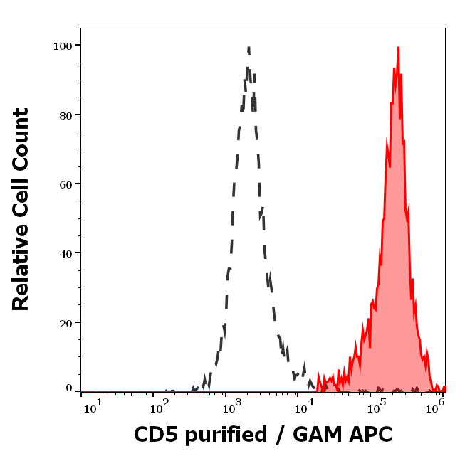
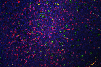
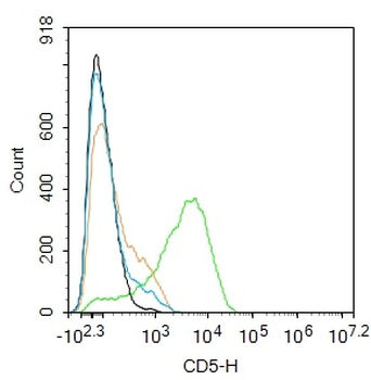
Separation of human CD5 positive lymphocytes (red-filled) from neutrophil granulocytes (black-dashed) in flow cytometry analysis (surface staining) of human peripheral whole blood stained using anti-human CD5 (L17F12) purified antibody (concentration in sample 2 μg/ml, GAM APC).
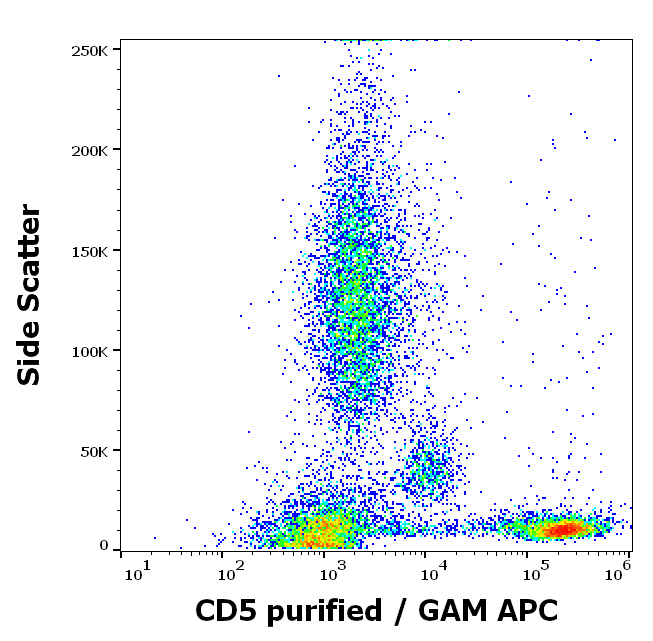
Flow cytometry surface staining pattern of human peripheral whole blood stained using anti-human CD5 (L17F12) purified antibody (concentration in sample 2 μg/ml, GAM APC).
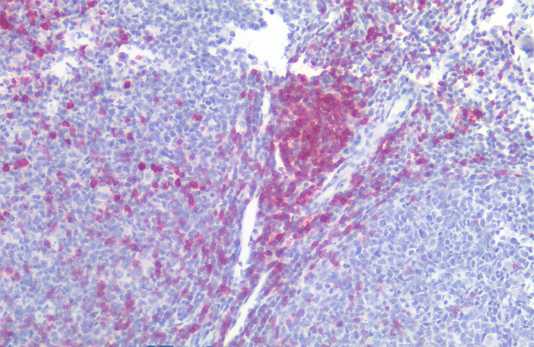
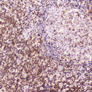
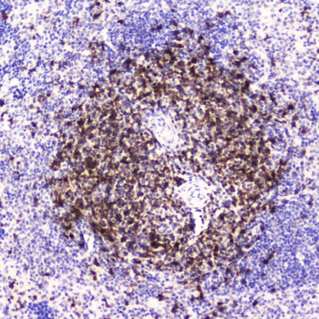
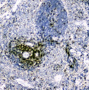
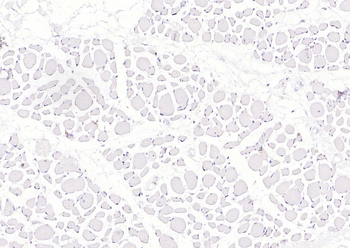
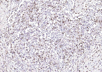
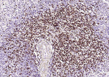
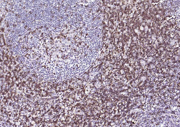
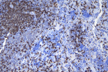
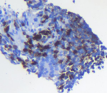
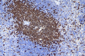
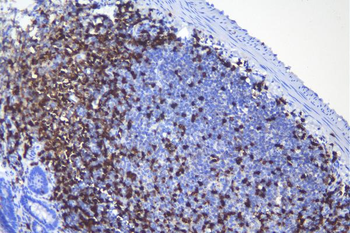
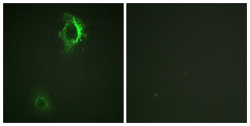
Immunohistochemistry staining of human tonsil (paraffin-embedded sections) with anti-CD5 (L17F12), 10 μg/ml.
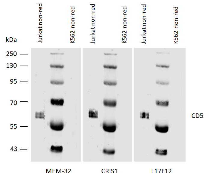
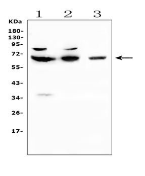
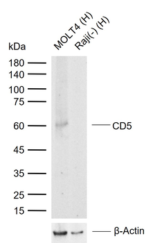
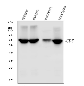
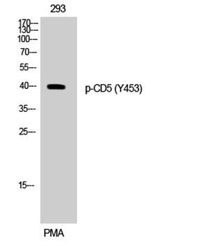
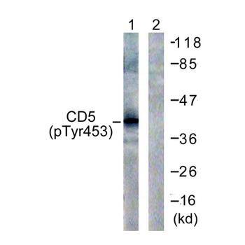
Western blotting analysis of human CD5 using mouse monoclonal antibodies MEM-32, CRIS1, and L17F12 on laurylmaltoside lysates of Jurkat cells and of K562 cells (negative control) under non-reducing conditions. Nitrocellulose membrane was probed with 2 µg/ml of mouse anti-CD5 monoclonal antibody followed by IRDye800-conjugated anti-mouse secondary antibody. CD5 was detected at approximately 62 kDa.
Documents Download
Datasheet
Product Information
Request a Document
Protocol Information
WB
Western Blot (IB, immunoblot)
IHC
Immunohistochemistry
FC
Flow Cytometry
ICC
Immunocytochemistry
IP
Immunoprecipitation
CD5 Antibody (orb154398)

- 0.0
Based on 0 reviews
Participating in our Biorbyt product reviews program enables you to support fellow scientists by sharing your firsthand experience with our products.
Login to Submit a ReviewAvailable Sizes
Select a size below
Free Secondary Antibody (20 ul)0/0
Please add an antibody product to your cart first.