You have no items in your shopping cart.
Description
Research Area
Immunology & Inflammation
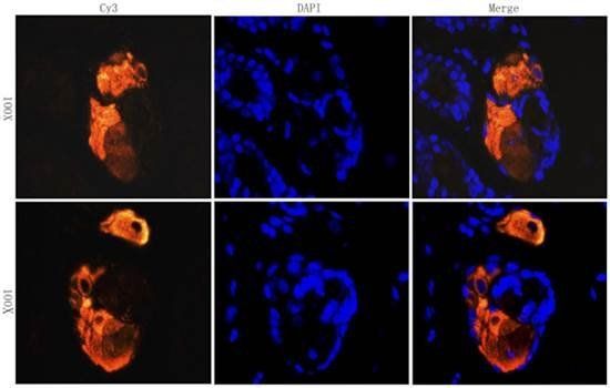
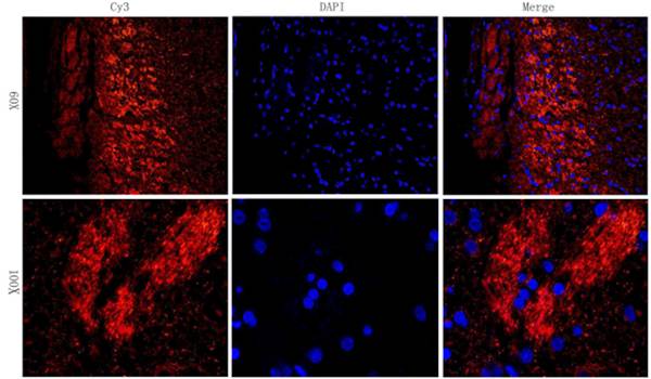
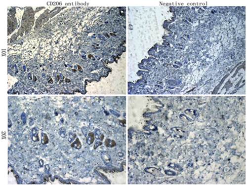
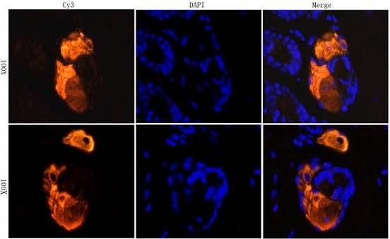
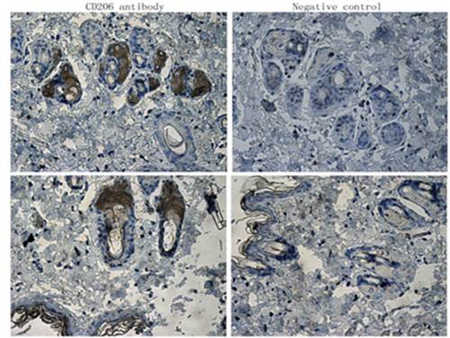
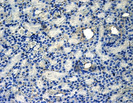
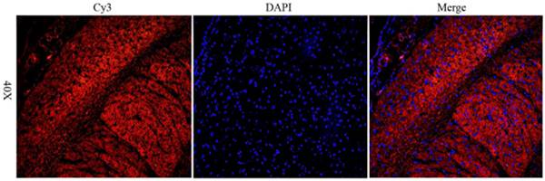
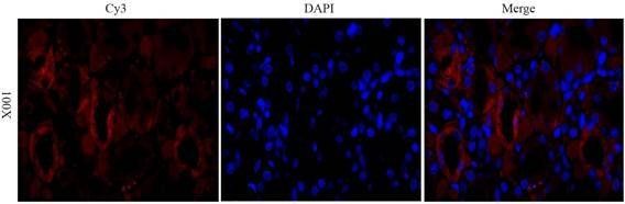
Images & Validation
−Item 1 of 3
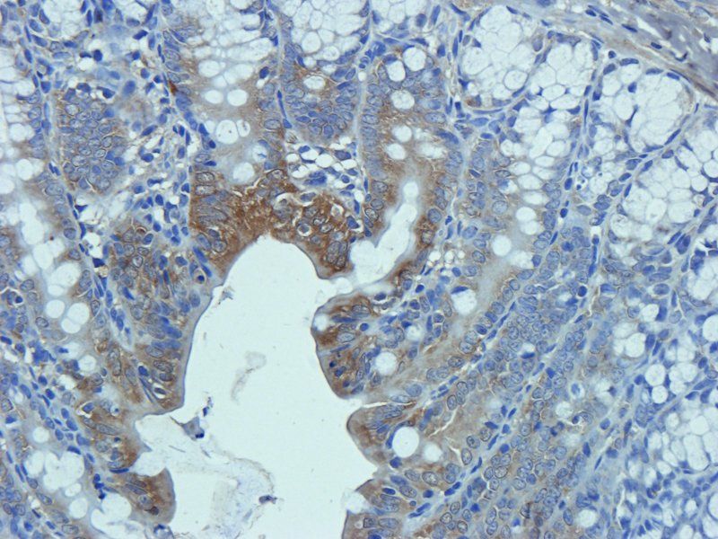
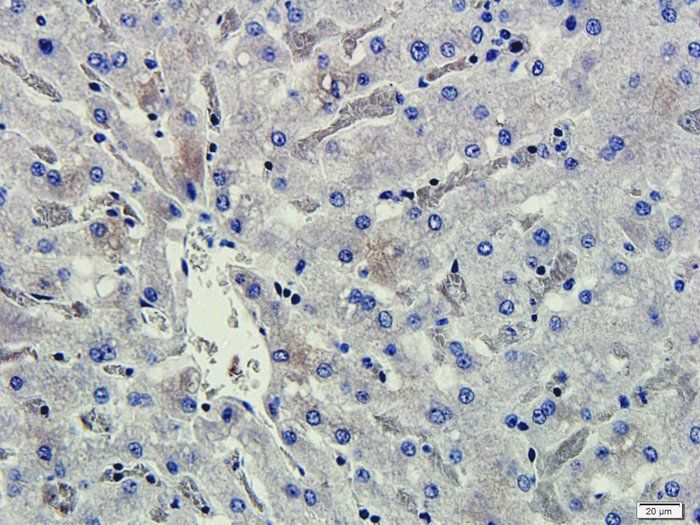
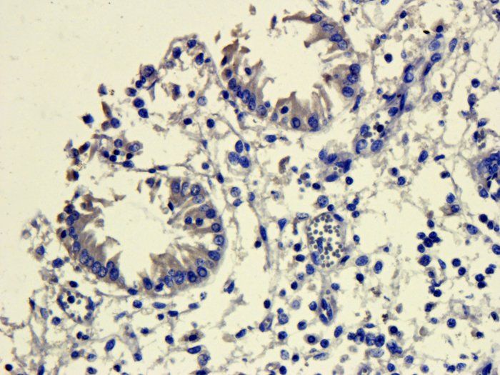
| Tested Applications | FA, FC, ICC, IHC-Fr, IP, WB |
|---|---|
| Reactivity | Human |
| Application Notes |
Key Properties
−| Antibody Type | Primary Antibody |
|---|---|
| Clonality | Monoclonal |
| Isotype | Mouse IgG1 kappa |
| Clone No. | 15-2 |
| Immunogen | Purified human mannose receptor |
| Target | CD206 |
| Purification | Purified by protein-A affinity chromatography. |
| Conjugation | Unconjugated |
Storage & Handling
−| Storage | Maintain refrigerated at 2-8°C for up to 2 weeks. For long term storage store at -20°C in small aliquots to prevent freeze-thaw cycles. |
|---|---|
| Buffer/Preservatives | Phosphate buffered saline (PBS), pH 7.4 |
| Concentration | 1 mg/ml |
| Expiration Date | 12 months from date of receipt. |
| Disclaimer | For research use only |
Alternative Names
−MMR, MRC1, CLEC13DL
Similar Products
−- Item 1 of 18
CD206 Rabbit Polyclonal Antibody [orb4941]
ELISA, ICC, IF, IHC-P, WB
Rabbit
Polyclonal
Unconjugated
200 μg, 1 mg, 100 μg - Item 1 of 8
CD206 Rabbit Polyclonal Antibody [orb180464]
ELISA, IHC-P
Human, Mouse, Porcine, Rat
Rabbit
Polyclonal
Unconjugated
100 μg - Item 1 of 5
CD206 Recombinant Rabbit Monoclonal Antibody [orb1610274]
ICC, IF, IHC-Fr, IHC-P
Human
Rabbit
Recombinant
Unconjugated
50 μl, 100 μl, 25 μl - Item 1 of 7
Mannose Receptor/MRC1 Rabbit Polyclonal Antibody [orb623873]
ELISA, IF, IHC, WB
Human, Monkey, Mouse, Rat
Rabbit
Polyclonal
Unconjugated
100 μg - Item 1 of 5
CD206 Rabbit Polyclonal Antibody [orb1294265]
IF, IHC, WB
Human, Mouse, Rat
Rabbit
Polyclonal
Unconjugated
100 μl, 25 μl


Quality Guarantee
Explore bioreagents carefree to elevate your research. All our products are rigorously tested for performance. If a product does not perform as described on its datasheet, our scientific support team will provide expert troubleshooting, a prompt replacement, or a refund. For full details, please see our Terms & Conditions and Buying Guide. Contact us at [email protected].
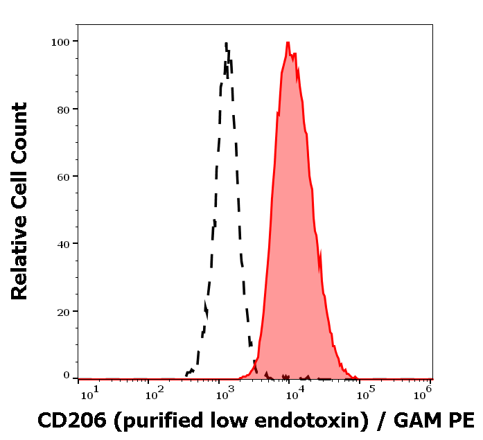
Separation of human CD206 positive dendritic cells differentiated upon monocyte stimulation (GM-CSF + IL-4) (red-filled) from non-stimulated lymphocytes (black-dashed) in flow cytometry analysis (surface staining) of human stimulated (GM-CSF + IL-4) peripheral blood mononuclear cells stained using anti-human CD206 (15-2) purified antibody (low endotoxin, concentration in sample 9 μg/ml), GAM PE.
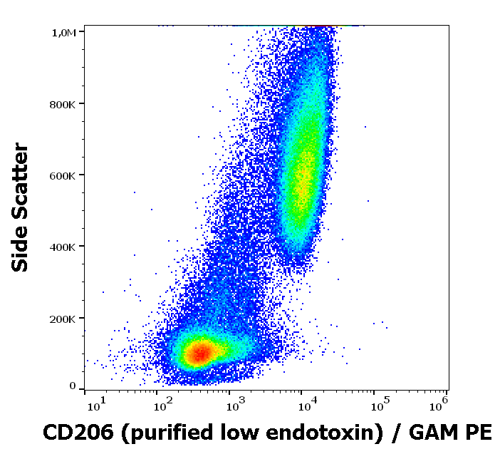
Flow cytometry surface staining pattern of human stimulated (GM-CSF + IL-4) peripheral blood mononuclear cells stained using anti-human CD206 (15-2) purified antibody (low endotoxin, concentration in sample 9 μg/ml), GAM PE.
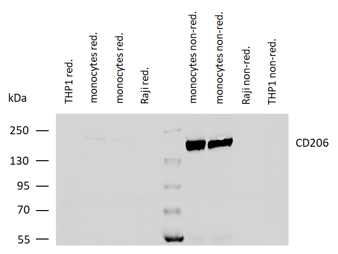
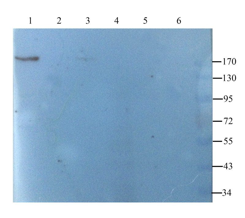
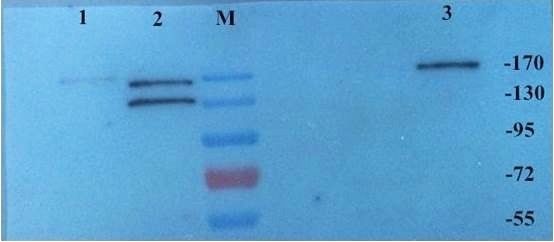
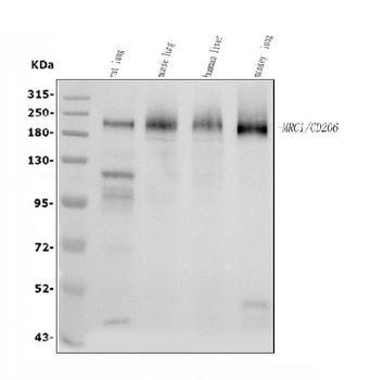
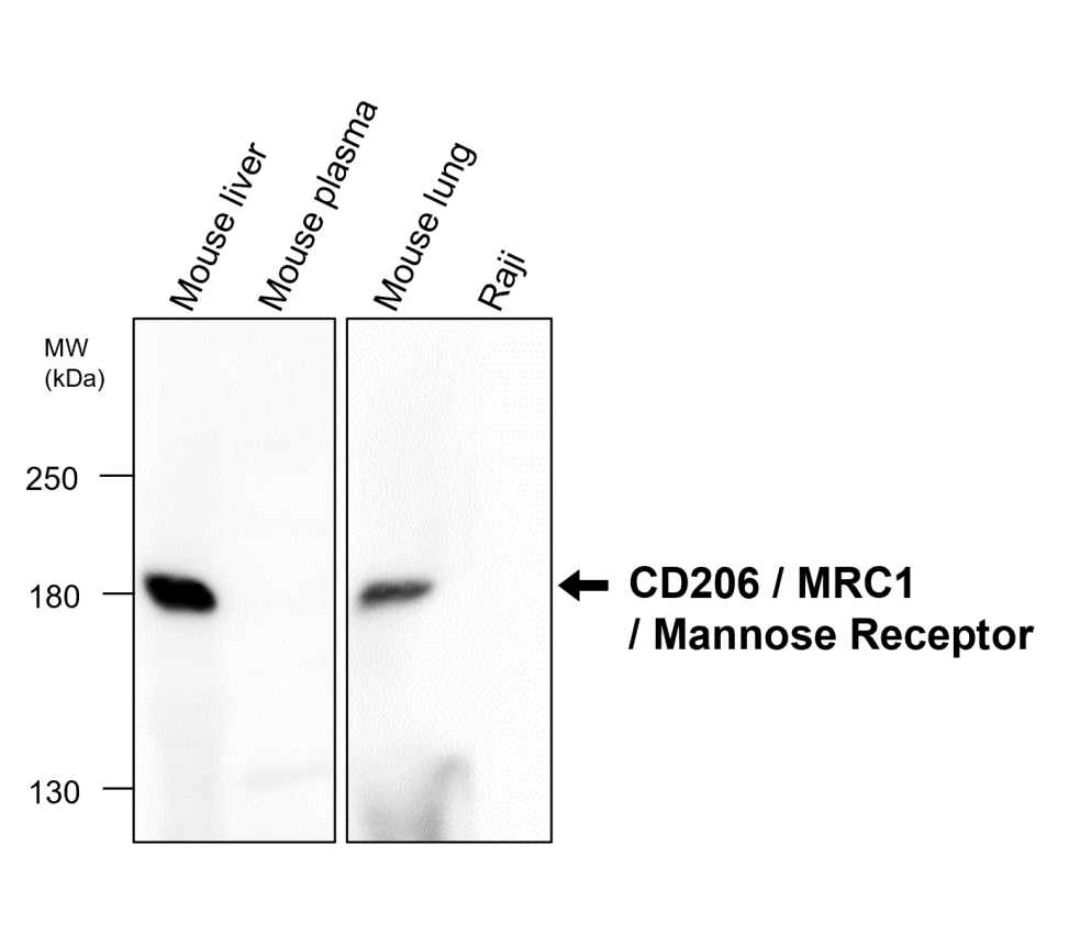
Western blotting analysis of human CD206 using mouse monoclonal antibody 15-2 on lysates of differentiated monocytes (two batches), and on THP-1 and Raji cells (negative controls) under reducing and non-reducing conditions. Nitrocellulose membrane was probed with 2 µg/ml of mouse monoclonal antibody followed by IRDye800-conjugated anti-mouse secondary antibody. CD206 was detected around 170 kDa.
Documents Download
Datasheet
Product Information
Request a Document
Protocol Information
WB
Western Blot (IB, immunoblot)
IHC-Fr
Immunohistochemistry Frozen
FC
Flow Cytometry
ICC
Immunocytochemistry
IP
Immunoprecipitation
CD206 Antibody (orb308746)

- 0.0
Based on 0 reviews
Participating in our Biorbyt product reviews program enables you to support fellow scientists by sharing your firsthand experience with our products.
Login to Submit a ReviewAvailable Sizes
Select a size below
Free Secondary Antibody (20 ul)0/0
Please add an antibody product to your cart first.