You have no items in your shopping cart.
Description
Research Area
Epigenetics & Chromatin, Molecular Biology
Images & Validation
−Item 1 of 3
| Tested Applications | ICC, IF, IHC, WB |
|---|---|
| Dilution Range | WB 1:500-2000 IHC 1:50-200 ICC/IF 1:50-200 |
| Reactivity | Human, Mouse, Rat |
Key Properties
−| Antibody Type | Primary Antibody |
|---|---|
| Host | Rabbit |
| Clonality | Monoclonal |
| Isotype | Rabbit IgG |
| Clone No. | BCGFHX0 |
| Immunogen | A synthesized peptide derived from human Histone H1.2 |
| Target | Histone H1.2 |
| Molecular Weight | 116 kDa |
| Purification | Affinity-chromatography |
| Conjugation | Unconjugated |
Storage & Handling
−| Storage | Maintain refrigerated at 2-8°C for up to 2 weeks. For long term storage store at -20°C in small aliquots to prevent freeze-thaw cycles. |
|---|---|
| Form/Appearance | Liquid |
| Buffer/Preservatives | Rabbit IgG in stabilizing components, phosphate buffered saline, pH 7.4, 150mM NaCl, 0.02% sodium azide and 50% glycerol. *This antibody is supplied in a stabilized formulation. Compatibility with conjugation reactions depends on the chemistry of the conjugation method used. For conjugation methods that are not compatible with the stabilizing components present in this formulation, a carrier-free antibody format is required. |
| Concentration | 0.5mg/ml |
| Expiration Date | 12 months from date of receipt. |
| Disclaimer | For research use only |
Alternative Names
−Histone deacetylase 11; HD11; 3.5.1.98; HDAC11
Quality Guarantee
Explore bioreagents carefree to elevate your research. All our products are rigorously tested for performance. If a product does not perform as described on its datasheet, our scientific support team will provide expert troubleshooting, a prompt replacement, or a refund. For full details, please see our Terms & Conditions and Buying Guide. Contact us at [email protected].
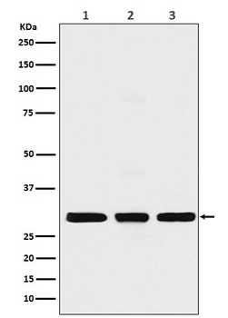
Western blot analysis of HIST1H1C using anti-HIST1H1C antibody (orb548408) in (1) MCF7 cell lysate (2) NIH/3T3 cell lysate; (3) C6 cell lysate. Electrophoresis was performed on a 5-20% SDS-PAGE gel at 70V (Stacking gel)/90V (Resolving gel) for 2-3 hours. The sample well of each lane was loaded with 50ug of sample under reducing conditions. After Electrophoresis, proteins were transferred to a Nitrocellulose membrane at 150mA for 50-90 minutes. Blocked the membrane with 5% Non-fat Milk/TBS for 1.5 hour at RT. The membrane was incubated with rabbit anti-HIST1H1C antigen affinity purified polyclonal antibody (Catalog # orb548408) at 0.5 ug/mL overnight at 4°C, then washed with TBS-0.1%Tween 3 times with 5 minutes each and probed with a goat anti-rabbit IgG-HRP secondary antibody at a dilution of 1:10000 for 1.5 hour at RT. The signal is developed using an Enhanced Chemiluminescent detection (ECL) kit (Catalog # orb90503) with Tanon 5200 system.
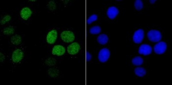
ICC staining Histone H1.2 in HeLa cells (green). The nuclear counter stain is DAPI (blue). Cells were fixed in paraformaldehyde, permeabilised with 0.25% Triton X100/PBS.
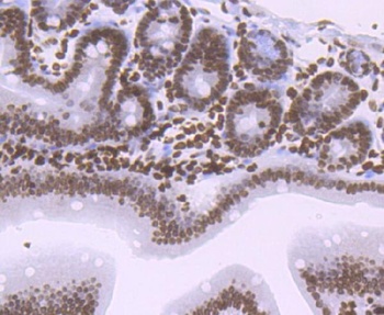
Immunohistochemical analysis of paraffin-embedded mouse colon tissue using anti-Histone H1.2 antibody. The section was pre-treated using heat mediated antigen retrieval with sodium citrate buffer (pH6) for 20 mins. Counter stained with hematoxylin.
Quick Database Links
Gene Symbol
Histone H1.2
UniProt
UniProt Details
− No UniProt data available
Documents Download
Datasheet
Product Information
Request a Document
Protocol Information
WB
Western Blot (IB, immunoblot)
IHC
Immunohistochemistry
IF
Immunofluorescence
ICC
Immunocytochemistry
Histone H1.2/H1 Rabbit Monoclonal Antibody (orb548408)

- 0.0
Based on 0 reviews
Participating in our Biorbyt product reviews program enables you to support fellow scientists by sharing your firsthand experience with our products.
Login to Submit a ReviewAvailable Sizes
Select a size below
Free Secondary Antibody (20 ul)0/0
Please add an antibody product to your cart first.