You have no items in your shopping cart.
Description
Research Area
Immunology & Inflammation
Images & Validation
−Item 1 of 2
| Tested Applications | IF, IHC |
|---|---|
| Dilution Range | Immunohistochemistry (Paraffin-embedded Section), 0.5-1μg/ml, Human Immunofluorescence, 5 μg/ml, Human |
| Reactivity | Human |
Related Conjugates & Formulations
−Key Properties
−| Antibody Type | Primary Antibody |
|---|---|
| Host | Rabbit |
| Clonality | Polyclonal |
| Isotype | Rabbit IgG |
| Immunogen | A synthetic peptide corresponding to a sequence in the middle region of human FDCSP. |
| Target | Follicular dendritic cell secreted peptide |
| Molecular Weight | 28 kDa |
| Purification | Immunogen affinity purified. |
| Conjugation | Unconjugated |
Storage & Handling
−| Storage | Maintain refrigerated at 2-8°C for up to 2 weeks. For long term storage store at -20°C in small aliquots to prevent freeze-thaw cycles. |
|---|---|
| Form/Appearance | Lyophilized |
| Buffer/Preservatives | Each vial contains 4mg Trehalose, 0.9mg NaCl, 0.2mg Na2HPO4, 0.05mg NaN3. |
| Concentration | Adding 0.2 ml of distilled water will yield a concentration of 500 μg/ml. |
| Expiration Date | 12 months from date of receipt. |
| Disclaimer | For research use only |
Alternative Names
−Follicular dendritic cell secreted peptide; FDC secreted protein; FDC-SP; FDCSP; C4orf7; UNQ733/PRO1419
Similar Products
−- Item 1 of 13
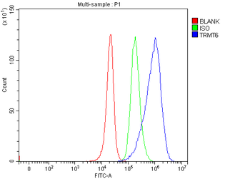
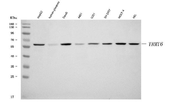
TRMT6 Rabbit Polyclonal Antibody [orb1291639]
ELISA, FC, ICC, IF, IHC, WB
Human
Rabbit
Polyclonal
Unconjugated
100 μg - Item 1 of 11
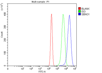
SBNO1 Rabbit Polyclonal Antibody [orb865568]
ELISA, FC, ICC, IF, IHC, WB
Human, Mouse, Rat
Rabbit
Polyclonal
Unconjugated
100 μg - Item 1 of 7
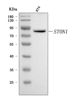
STON1 Rabbit Polyclonal Antibody [orb1676526]
ELISA, FC, IF, IHC, WB
Human
Rabbit
Polyclonal
Unconjugated
100 μg - Item 1 of 6
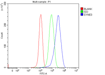
Nesprin3/SYNE3 Rabbit Polyclonal Antibody [orb1676431]
ELISA, FC, IHC, WB
Human, Mouse
Rabbit
Polyclonal
Unconjugated
100 μg - Item 1 of 5
NUDCD3 Rabbit Polyclonal Antibody [orb1743791]
ELISA, FC, ICC, IF, IHC, WB
Human
Rabbit
Polyclonal
Unconjugated
100 μg


Quality Guarantee
Explore bioreagents carefree to elevate your research. All our products are rigorously tested for performance. If a product does not perform as described on its datasheet, our scientific support team will provide expert troubleshooting, a prompt replacement, or a refund. For full details, please see our Terms & Conditions and Buying Guide. Contact us at [email protected].

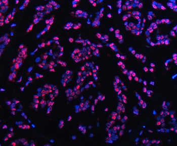
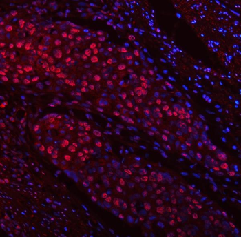
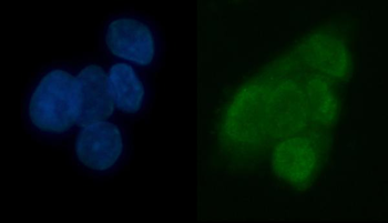
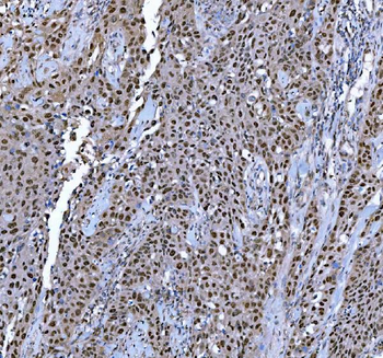
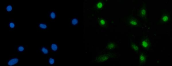
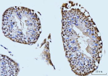
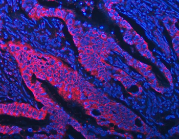
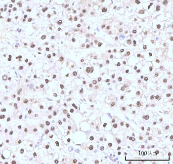
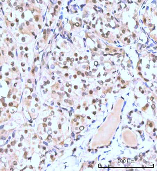
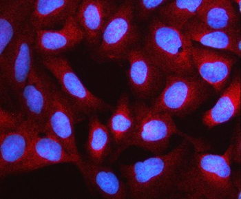
IF analysis of FDCSP using anti-FDCSP antibody. FDCSP was detected in a paraffin-embedded section of human tonsil tissue. Heat mediated antigen retrieval was performed in EDTA buffer (pH8.0, epitope retrieval solution). The tissue section was blocked with 10% goat serum. The tissue section was then incubated with 5 µg/mL rabbit anti-FDCSP Antibody overnight at 4°C. FITC Conjugated Goat Anti-Rabbit IgG was used as secondary antibody at 1:500 dilution and incubated for 30 minutes at 37°C. The section was counterstained with DAPI. Visualize using a fluorescence microscope and filter sets appropriate for the label used.
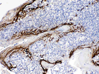
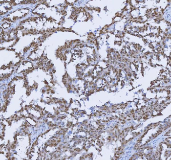
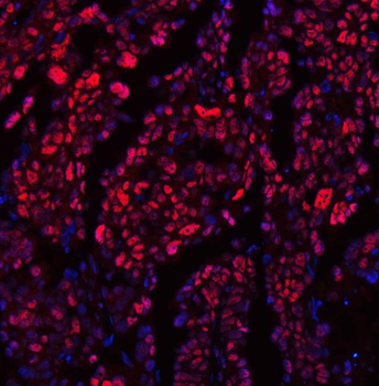
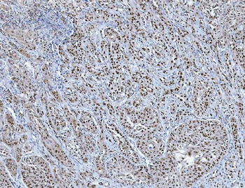
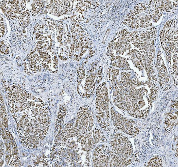
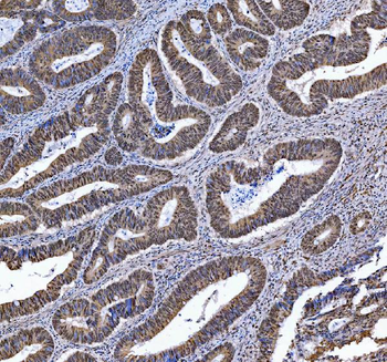
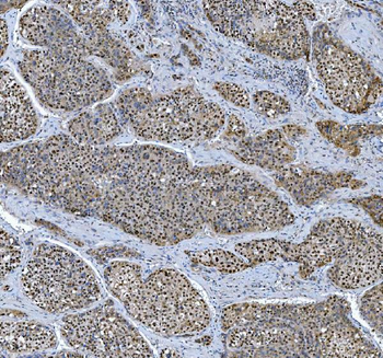
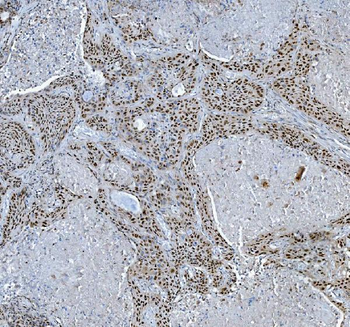
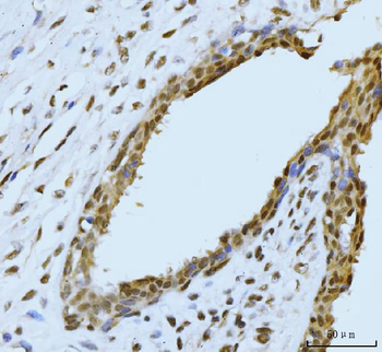
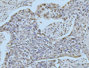
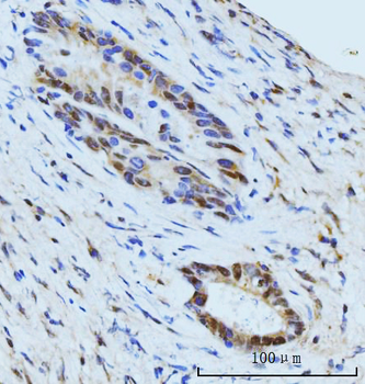
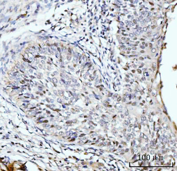
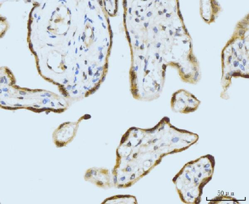
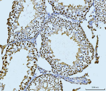
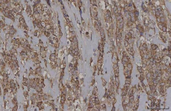
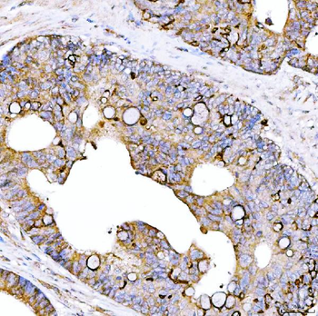
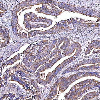
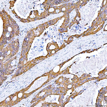
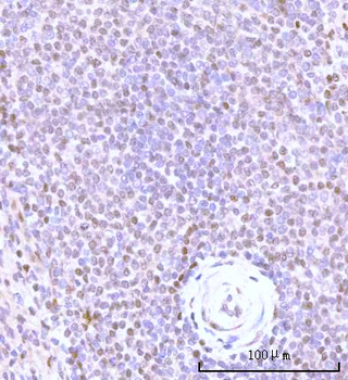
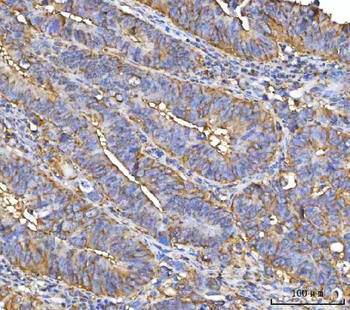
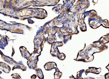
IHC analysis of FDCSP using anti-FDCSP antibody. FDCSP was detected in paraffin-embedded section of human tonsil tissues. Heat mediated antigen retrieval was performed in citrate buffer (pH6, epitope retrieval solution) for 20 mins. The tissue section was blocked with 10% goat serum. The tissue section was then incubated with 1 µg/ml rabbit anti-FDCSP Antibody overnight at 4°C. Biotinylated goat anti-rabbit IgG was used as secondary antibody and incubated for 30 minutes at 37°C. The tissue section was developed using Strepavidin-Biotin-Complex (SABC) with DAB as the chromogen.
Quick Database Links
Gene Symbol
Follicular dendritic cell secreted peptide
UniProt
UniProt Details
− No UniProt data available
Documents Download
Datasheet
Product Information
Request a Document
Protocol Information
IHC
Immunohistochemistry
IF
Immunofluorescence
FDCSP Rabbit Polyclonal Antibody (orb18784)

- 0.0
Based on 0 reviews
Participating in our Biorbyt product reviews program enables you to support fellow scientists by sharing your firsthand experience with our products.
Login to Submit a ReviewAvailable Sizes
Select a size below
Free Secondary Antibody (20 ul)0/0
Please add an antibody product to your cart first.