You have no items in your shopping cart.
Description
Research Area
Cancer Biology, Cell Biology, Disease Biomarkers, Immunology & Inflammation, Stem Cell & Developmental Biology
Images & Validation
−Item 1 of 5
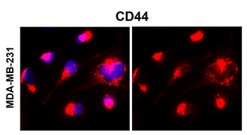
| Tested Applications | IF, IHC, WB |
|---|---|
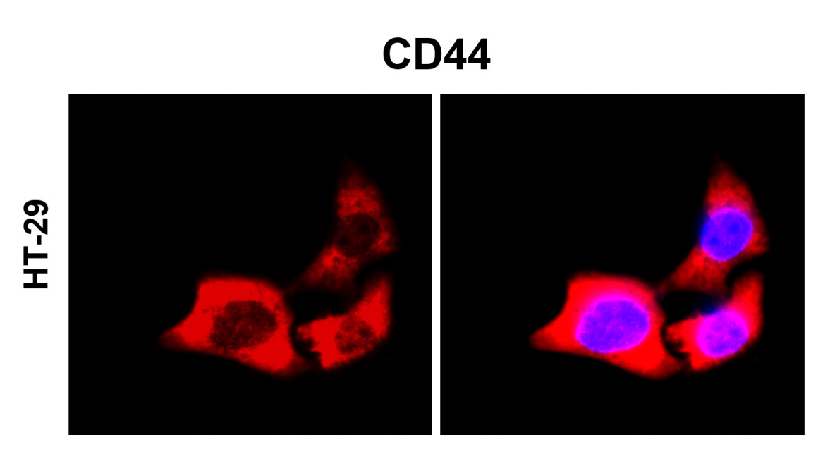
| Dilution Range | Western blot, 0.1-0.5μg/ml, Human, Rat Immunohistochemistry (Paraffin-embedded Section), 0.5-1μg/ml, Human Immunofluorescence, 2μg/ml, Human, Rat |
| Reactivity | Human, Rat |
Related Conjugates & Formulations
−Key Properties
−| Antibody Type | Primary Antibody |
|---|---|
| Host | Rabbit |
| Clonality | Polyclonal |
| Isotype | Rabbit IgG |
| Immunogen | A synthetic peptide corresponding to a sequence at the N-terminus of human CD44, different from the related mouse and rat sequences by two amino acids. |
| Target | CD44 antigen |
| Molecular Weight | 82 kDa |
| Purification | Immunogen affinity purified. |
| Conjugation | Unconjugated |
Storage & Handling
−| Storage | Maintain refrigerated at 2-8°C for up to 2 weeks. For long term storage store at -20°C in small aliquots to prevent freeze-thaw cycles. |
|---|---|
| Form/Appearance | Lyophilized |
| Buffer/Preservatives | Each vial contains antibody formulated with stabilizing components, 0.9 mg NaCl, 0.2 mg Na2HPO4, and 0.05 mg NaN3. *This antibody is supplied in a stabilized formulation. Compatibility with conjugation reactions depends on the chemistry of the conjugation method used. For conjugation methods that are not compatible with the stabilizing components present in this formulation, a carrier-free antibody format is required. |
| Concentration | Adding 0.2 ml of distilled water will yield a concentration of 500 μg/ml. |
| Expiration Date | 12 months from date of receipt. |
| Disclaimer | For research use only |
Alternative Names
−CD44 antigen; CDw44; Epican; Extracellular matrix receptor III; ECMR-III; GP90 lymphocyte homing/adhesion receptor; HUTCH-I; Heparan sulfate proteoglycan; Hermes antigen; Hyaluronate receptor; Phagocytic glycoprotein 1; PGP-1; Phagocytic glycoprotein I; PGP-I; CD44; CD44; LHR, MDU2, MDU3, MIC4
Similar Products
−- Item 1 of 9
CD44 Rabbit Polyclonal Antibody [orb259645]
ELISA, IHC-P, WB
Human, Mouse, Porcine, Rat
Rabbit
Polyclonal
Unconjugated
100 μg - Item 1 of 8
CD44 Rabbit Polyclonal Antibody [orb402179]
ELISA, FC, ICC, IF, IHC, IHC-Fr, WB
Human, Mouse, Rat
Rabbit
Polyclonal
Unconjugated
100 μg - Item 1 of 8
CD44 Rabbit Polyclonal Antibody [orb1294318]
IF, IHC, WB
Human, Mouse, Rat
Rabbit
Polyclonal
Unconjugated
100 μl, 25 μl - Item 1 of 7
CD44 Rabbit Polyclonal Antibody [orb1972567]
ELISA, FC, ICC, IF, IHC, KO/KD Validated, WB
Human, Mouse, Rat
Rabbit
Polyclonal
Unconjugated
100 μg - Item 1 of 7
CD44 Polyclonal Antibody [orb1414374]
IF, IHC-P, WB
Human, Mouse, Rat
Rabbit
Polyclonal
Unconjugated
100 μl
Quality Guarantee
Explore bioreagents carefree to elevate your research. All our products are rigorously tested for performance. If a product does not perform as described on its datasheet, our scientific support team will provide expert troubleshooting, a prompt replacement, or a refund. For full details, please see our Terms & Conditions and Buying Guide. Contact us at [email protected].

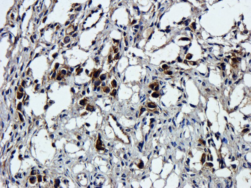
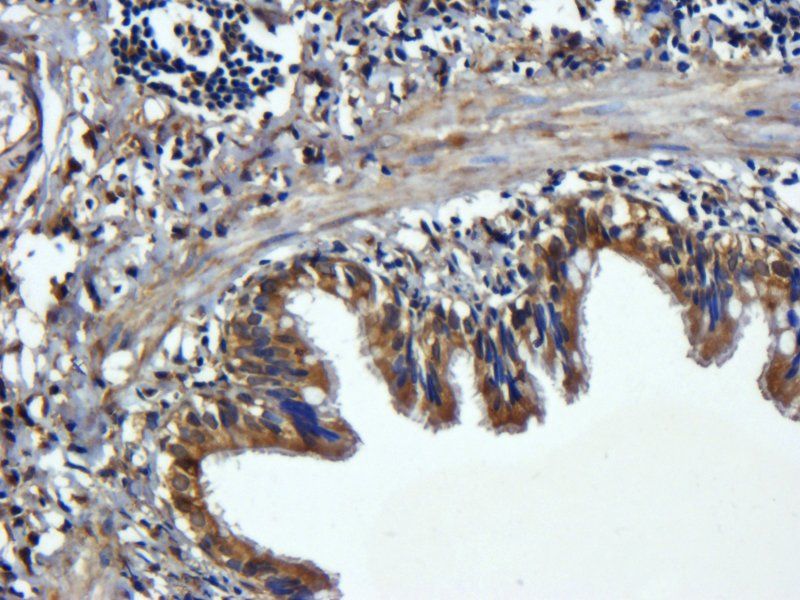
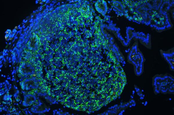
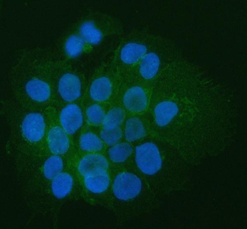
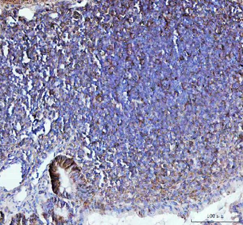
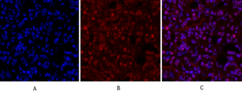
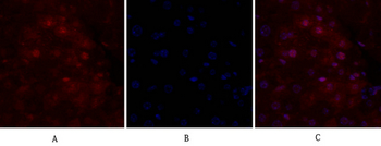
IF analysis of CD44 using anti-CD44 antibody. CD44 was detected in paraffin-embedded section of human tonsil tissues. Heat mediated antigen retrieval was performed in citrate buffer (pH6, epitope retrieval solution) for 20 mins. The tissue section was blocked with 10% goat serum. The tissue section was then incubated with 1 µg/mL rabbit anti-CD44 Antibody overnight at 4°C. DyLight®488 Conjugated Goat Anti-Rabbit IgG was used as secondary antibody at 1:100 dilution and incubated for 30 minutes at 37°C. The section was counterstained with DAPI. Visualize using a fluorescence microscope and filter sets appropriate for the label used.
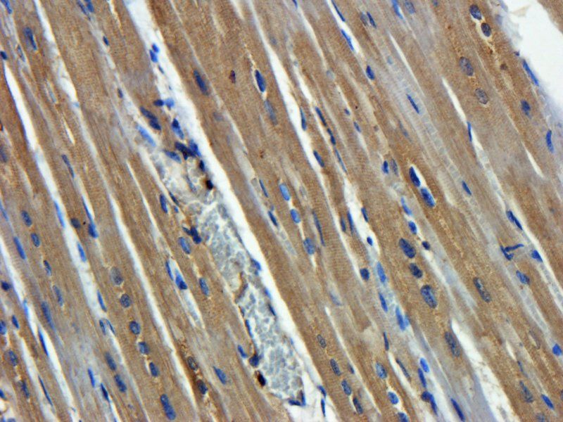
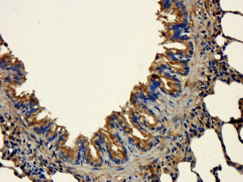
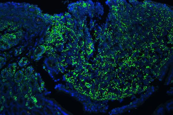
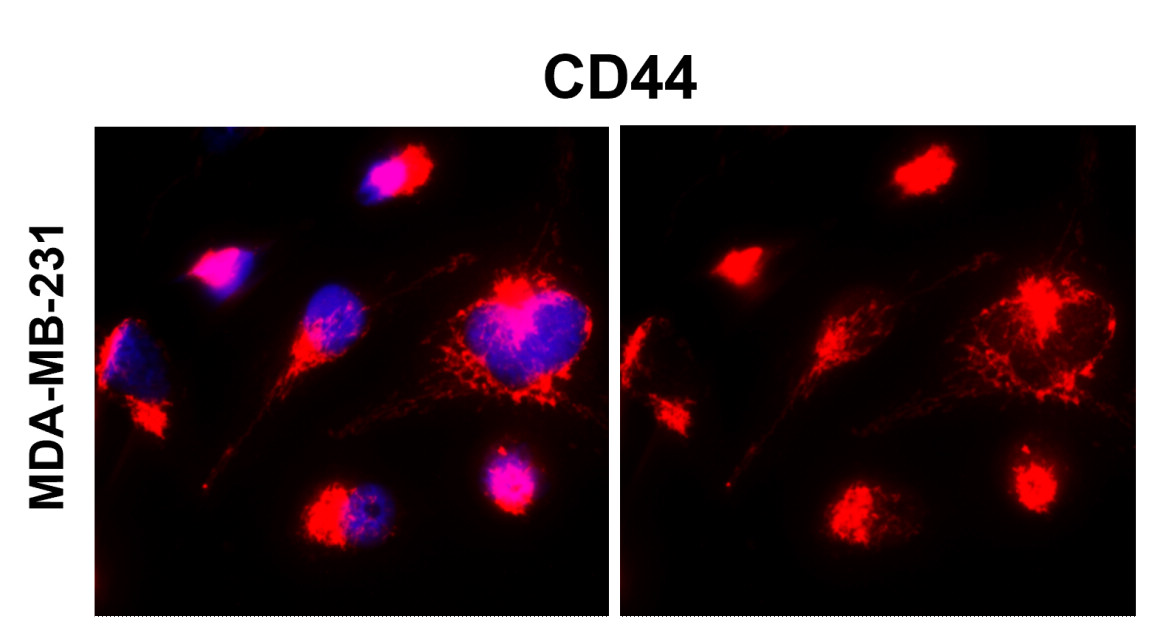
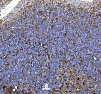
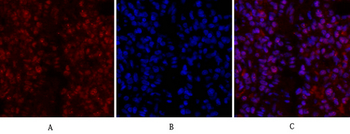
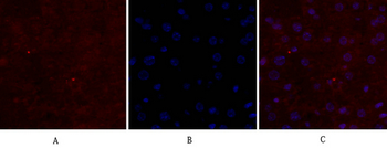
IF analysis of CD44 using anti-CD44 antibody. CD44 was detected in paraffin-embedded section of rat lymphaden tissues. Heat mediated antigen retrieval was performed in citrate buffer (pH6, epitope retrieval solution) for 20 mins. The tissue section was blocked with 10% goat serum. The tissue section was then incubated with 1 µg/mL rabbit anti-CD44 Antibody overnight at 4°C. DyLight®488 Conjugated Goat Anti-Rabbit IgG was used as secondary antibody at 1:100 dilution and incubated for 30 minutes at 37°C. The section was counterstained with DAPI. Visualize using a fluorescence microscope and filter sets appropriate for the label used.
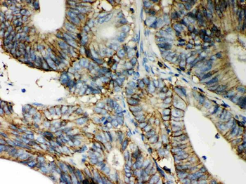
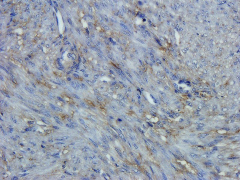
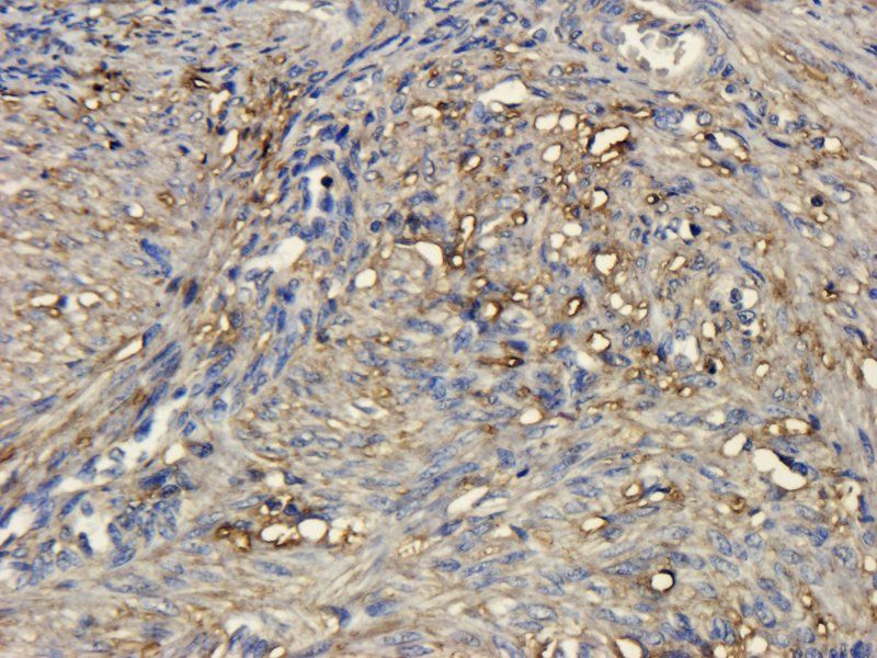
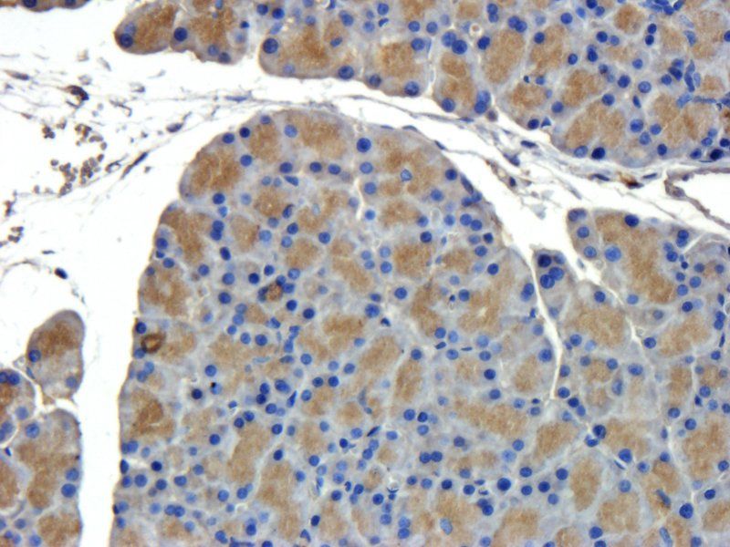
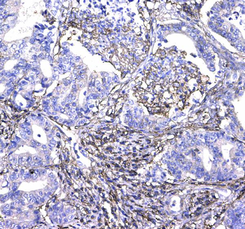
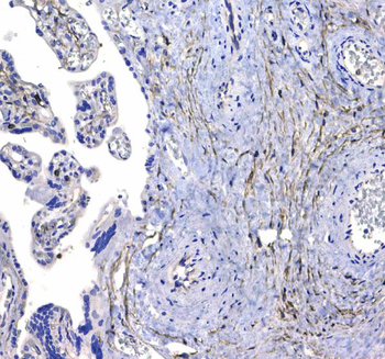
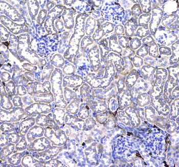
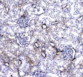
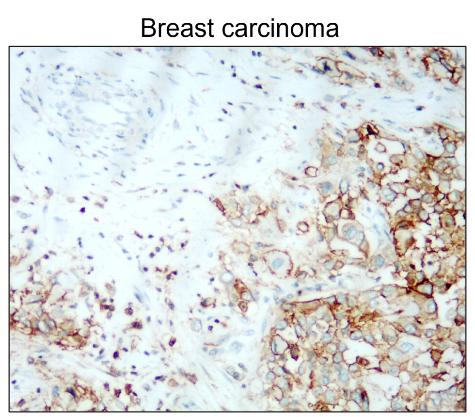
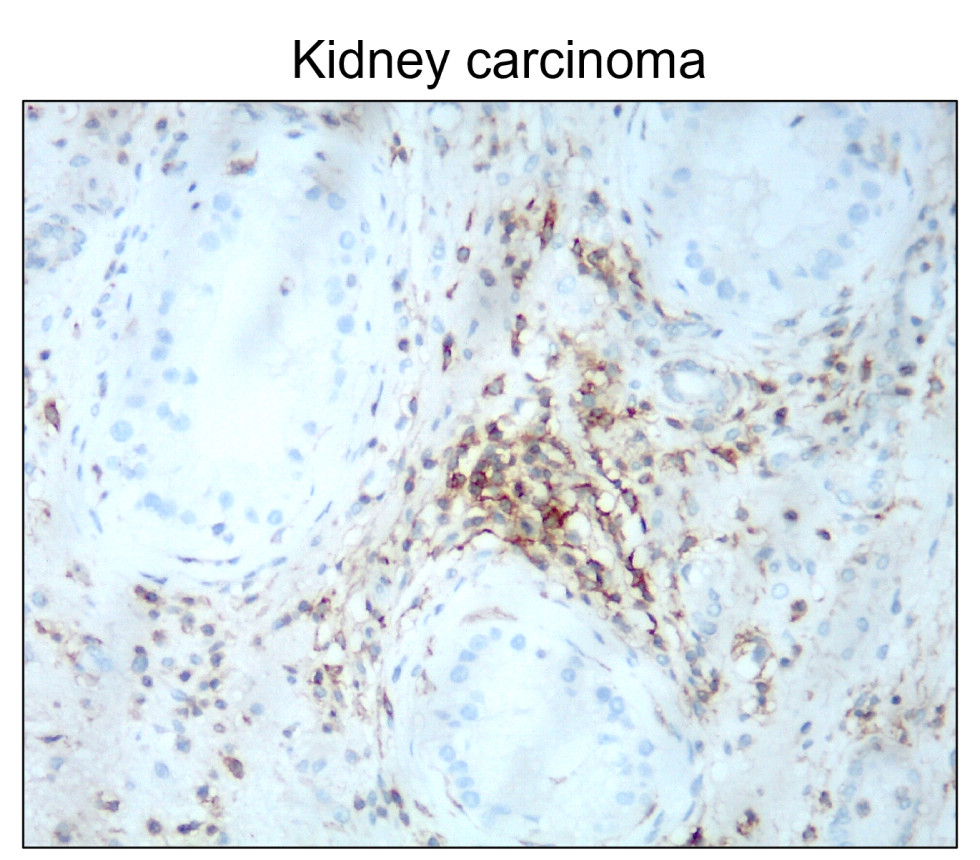
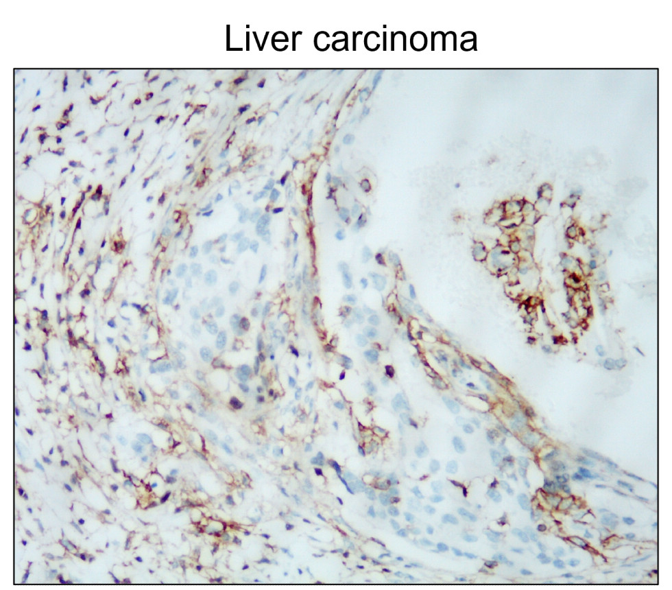
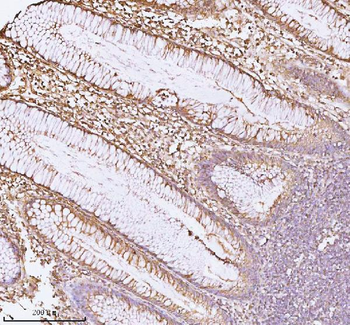
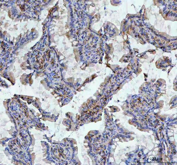
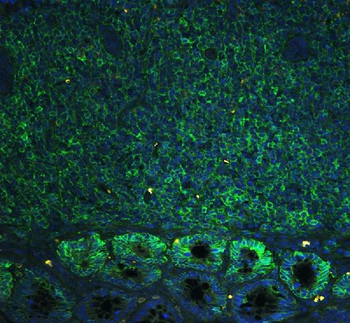
IHC analysis of CD44 using anti-CD44 antibody. CD44 was detected in a paraffin-embedded section of Human Intestinal Cancer tissue. Heat mediated antigen retrieval was performed in EDTA buffer (pH8.0, epitope retrieval solution). The tissue section was blocked with 10% goat serum. The tissue section was then incubated with 1 µg/ml rabbit anti-CD44 Antibody overnight at 4°C. Peroxidase Conjugated Goat Anti-rabbit IgG was used as secondary antibody and incubated for 30 minutes at 37°C. The tissue section was developed using HRP Conjugated Rabbit IgG Super Vision Assay Kit with DAB as the chromogen.

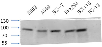
Western blot analysis of CD44 using anti-CD44 antibody. Electrophoresis was performed on a 5-20% SDS-PAGE gel at 70V (Stacking gel) / 90V (Resolving gel) for 2-3 hours. The sample well of each lane was loaded with 30 ug of sample under reducing conditions. After electrophoresis, proteins were transferred to a nitrocellulose membrane at 150 mA for 50-90 minutes. Blocked the membrane with 5% milk in PBS/0.05% Tween-20 (5% milk/PBS/Tw) for 1.5 hour at RT. The membrane was incubated with rabbit anti-CD44 antibody at 1 ug/mL in 5% milk/PBS/0.05% Tween 20 overnight at 4℃, then washed with TBS-0.1% Tween 3 times with 5 minutes each and probed with a goat anti-rabbit antibody conjugated with HRP at 1:5000 in 5% milk/PBS/Tw at 4℃ for 12 hours. The signal is developed using an SuperSignal West Pico Chemiluminescent Substrate. A specific band was detected for EGFR at approximately 20 kDa. The expected band size for EGFR is at 81 kDa.
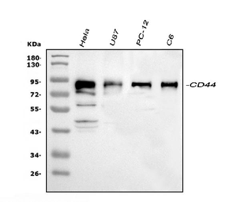
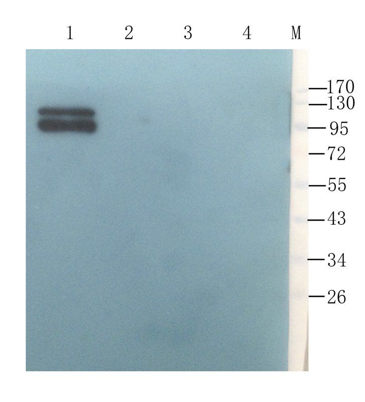
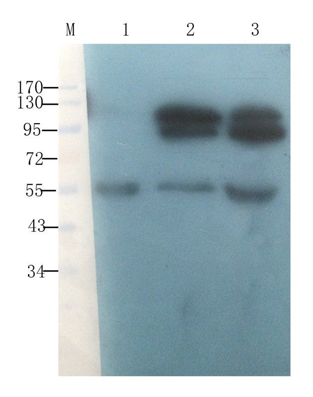
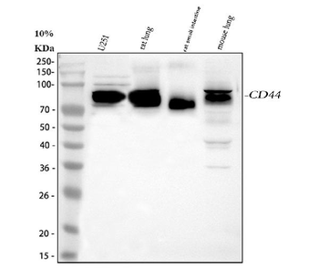
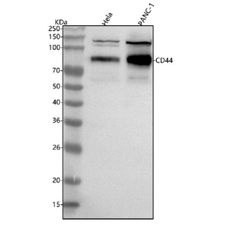
Western blot analysis of CD44 using anti-CD44 antibody. Electrophoresis was performed on a 5-20% SDS-PAGE gel at 70V (Stacking gel) / 90V (Resolving gel) for 2-3 hours. The sample well of each lane was loaded with 30 ug of sample under reducing conditions. Lane 1: human Hela whole cell lysates, Lane 2: human U87 whole cell lysates, Lane 3: rat PC-12 whole cell lysates, Lane 4: rat C6 whole cell lysates. After electrophoresis, proteins were transferred to a nitrocellulose membrane at 150 mA for 50-90 minutes. Blocked the membrane with 5% non-fat milk/TBS for 1.5 hour at RT. The membrane was incubated with rabbit anti-CD44 antigen affinity purified polyclonal antibody at 0.5 µg/mL overnight at 4°C, then washed with TBS-0.1% Tween 3 times with 5 minutes each and probed with a goat anti-rabbit IgG-HRP secondary antibody at a dilution of 1:5000 for 1.5 hour at RT. The signal is developed using an Enhanced Chemiluminescent detection (ECL) kit with Tanon 5200 system. A specific band was detected for CD44 at approximately 82 kDa. The expected band size for CD44 is at 82 kDa.
Quick Database Links
Gene Symbol
CD44 antigen
UniProt
UniProt Details
− No UniProt data available
Documents Download
Datasheet
Product Information
Request a Document
Protocol Information
WB
Western Blot (IB, immunoblot)
IHC
Immunohistochemistry
IF
Immunofluorescence
CD44 Rabbit Polyclonal Antibody (orb251516)

- 0.0
Based on 0 reviews
Participating in our Biorbyt product reviews program enables you to support fellow scientists by sharing your firsthand experience with our products.
Login to Submit a ReviewAvailable Sizes
Select a size below
Free Secondary Antibody (20 ul)0/0
Please add an antibody product to your cart first.