You have no items in your shopping cart.
Featured
Description
Research Area
Stem Cell & Developmental Biology
Images & Validation
−Item 1 of 2
| Tested Applications | IF, WB |
|---|---|
| Dilution Range | WB: 1:500-1000 |
| Reactivity | Human, Monkey, Mouse, Rat |
Key Properties
−| Antibody Type | Primary Antibody |
|---|---|
| Host | Rabbit |
| Clonality | Polyclonal |
| Immunogen | KLH-conjugated synthetic peptide encompassing a sequence within the center region of human CATSPER1. The exact sequence is proprietary. |
| Target | CATSPER1 |
| Purification | The antibody was purified by immunogen affinity chromatography. |
| Conjugation | Unconjugated |
Storage & Handling
−| Storage | Maintain refrigerated at 2-8°C for up to 2 weeks. For long term storage store at -20°C in small aliquots to prevent freeze-thaw cycles. |
|---|---|
| Form/Appearance | Liquid |
| Buffer/Preservatives | 0.42% Potassium phosphate, 0.87% Sodium chloride, pH 7.3, 30% glycerol, and 0.01% sodium azide. |
| Expiration Date | 12 months from date of receipt. |
| Disclaimer | For research use only |
Alternative Names
−Cation channel sperm-associated protein 1; CatSper1; hCatSper
Similar Products
−- Item 1 of 5
CATSPER Rabbit Polyclonal Antibody [orb704210]
IF, IHC-Fr, IHC-P, WB
Mouse, Rat
Human, Mouse, Rat
Rabbit
Polyclonal
Unconjugated
50 μl, 100 μl, 200 μl - Item 1 of 1
- Item 1 of 1
CATSPER Rabbit Polyclonal Antibody [orb420631]
WB
Rat
Mouse
Rabbit
Polyclonal
Unconjugated
50 μl, 100 μl, 200 μl 


Quality Guarantee
Explore bioreagents carefree to elevate your research. All our products are rigorously tested for performance. If a product does not perform as described on its datasheet, our scientific support team will provide expert troubleshooting, a prompt replacement, or a refund. For full details, please see our Terms & Conditions and Buying Guide. Contact us at [email protected].
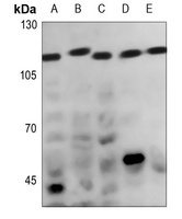
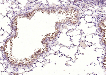
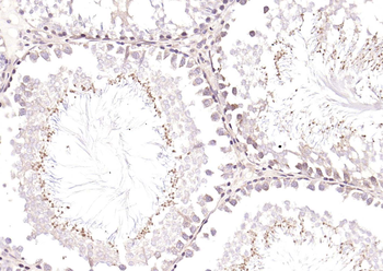
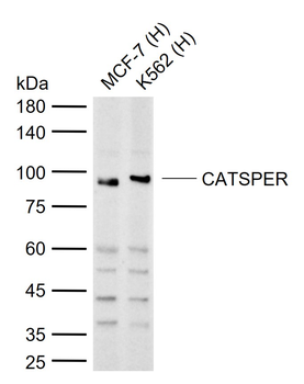
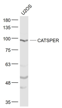
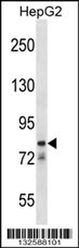
Western blot analysis of CATSPER1 expression in Hela (A), DLD (B), A375 (C), mouse testis (D), rat testis (E) whole cell lysates. (Predicted band size: 90 kD; Observed band size: 110 kD)
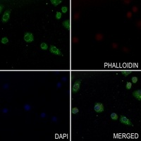
Immunofluorescent analysis of CATSPER1 staining in SGC7901 cells. Formalin-fixed cells were permeabilized with 0.1% Triton X-100 in TBS for 5-10 minutes and blocked with 3% BSA-PBS for 30 minutes at room temperature. Cells were probed with the primary antibody in 3% BSA-PBS and incubated overnight at 4 °C in a hidified chamber. Cells were washed with PBST and incubated with a AF488-conjugated secondary antibody (green) in PBS at room temperature in the dark. Phalloidin - AF594 was used to stain Actin filaments (red). DAPI was used to stain the cell nuclei (blue).
Quick Database Links
UniProt Details
− No UniProt data available
NCBI Gene Details
− No NCBI Gene data available
Documents Download
Datasheet
Product Information
Request a Document
Protocol Information
WB
Western Blot (IB, immunoblot)
IF
Immunofluorescence
Filter by Applications
Filter by Species
Jing Gu 1, Ning Zhang 1, Xiao Jiang 1, Lei Zhu 1, Yixia Lou 1, Shengqi Sun 1, Li Yin 1 2, Jinyi Liu 1 The Olfactory Receptor Olfr25 Mediates Sperm Dysfunction Induced by Low-Dose Bisphenol A through the CatSper-Ca2+ Signaling Pathway Toxics, 12, 442 (2024)
Applications
WB
Reactivity
Mouse
CATSPER1 Rabbit Polyclonal Antibody (orb234792)

- 0.0
Based on 0 reviews
Participating in our Biorbyt product reviews program enables you to support fellow scientists by sharing your firsthand experience with our products.
Login to Submit a ReviewAvailable Sizes
Select a size below
Choose Conjugation or Carrier Free Version
Free Secondary Antibody (20 ul)0/0
Please add an antibody product to your cart first.