Queen's Award Received in 2021 ISO 9001 Certified Delivered over 1,000,000 bio-reagents to life science researchers Trusted by Life Science Communities
Cart summary

You have no items in your shopping cart.

Cathepsin G/CTSG Rabbit Polyclonal Antibody (Cy3)

SKU: orb2573925

Description

Anti-Cathepsin G/CTSG Antibody. Tested in WB applications. This antibody reacts with Human, Rat.

Research Area

Immunology & Inflammation

Images & Validation

Tested ApplicationsFC
Dilution RangeFlow Cytometry, 1-3μg/1x10^6 cells
ReactivityHuman, Rat

Related Conjugates & Formulations

Key Properties

Antibody TypePrimary Antibody
HostRabbit
ClonalityPolyclonal
IsotypeRabbit IgG
ImmunogenE.coli-derived human Cathepsin G recombinant protein (Position: A76-L255). Human Cathepsin G shares 66% amino acid (aa) sequence identity with mouse Cathepsin G.
TargetCathepsin G
Molecular Weight29 kDa
PurificationImmunogen affinity purified.
ConjugationCy3

Storage & Handling

StorageAt -20°C for one year from date of receipt. Avoid repeated freezing and thawing. Protect from light.
Form/AppearanceLiquid
Buffer/PreservativesEach vial contains 50% glycerol, 0.9% NaCl, 0.2% Na2HPO4, 0.02% NaN3.
Expiration Date12 months from date of receipt.
DisclaimerFor research use only

Alternative Names

Cathepsin G; CG; 3.4.21.20; CTSG

Similar Products

Quality Guarantee

Quality Guarantee

Explore bioreagents carefree to elevate your research. All our products are rigorously tested for performance. If a product does not perform as described on its datasheet, our scientific support team will provide expert troubleshooting, a prompt replacement, or a refund. For full details, please see our Terms & Conditions and Buying Guide. Contact us at [email protected].

Cathepsin G/CTSG Rabbit Polyclonal Antibody (Cy3)

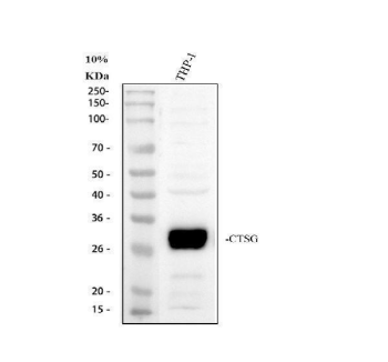
Western blot analysis of Cathepsin G using anti-Cathepsin G antibody. Electrophoresis was performed on a 10% SDS-PAGE gel at 80V (Stacking gel) / 120V (Resolving gel) for 2 hours. The sample well of each lane was loaded with 30 ug of sample under reducing conditions. Lane 1: human THP-1 whole cell lysates. After electrophoresis, proteins were transferred to a nitrocellulose membrane at 150 mA for 50-90 minutes. Blocked the membrane with 5% non-fat milk/TBS for 1.5 hour at RT. The membrane was incubated with rabbit anti-Cathepsin G antigen affinity purified polyclonal antibody at 0.5 μg/mL overnight at 4°C, then washed with TBS-0.1%Tween 3 times with 5 minutes each and probed with a goat anti-rabbit IgG-HRP secondary antibody at a dilution of 1:5000 for 1.5 hour at RT. The signal is developed using an ECL Plus Western Blotting Substrate with Tanon 5200 system. A specific band was detected for Cathepsin G at approximately 29 kDa. The expected band size for Cathepsin G is at 29 kDa.

Cathepsin G/CTSG Rabbit Polyclonal Antibody (Cy3)

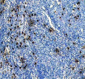
IHC analysis of Cathepsin G using anti-Cathepsin G antibody. Cathepsin G was detected in a paraffin-embedded section of human tonsil tissue. Heat mediated antigen retrieval was performed in EDTA buffer (pH 8.0, epitope retrieval solution). The tissue section was blocked with 10% goat serum. The tissue section was then incubated with 2 μg/ml rabbit anti-Cathepsin G Antibody (PB9049) overnight at 4°C. Peroxidase Conjugated Goat Anti-rabbit IgG was used as secondary antibody and incubated for 30 minutes at 37°C. The tissue section was developed using HRP Conjugated Rabbit IgG Super Vision Assay Kit with DAB as the chromogen.

Cathepsin G/CTSG Rabbit Polyclonal Antibody (Cy3)

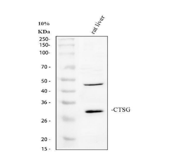
Western blot analysis of Cathepsin G using anti-Cathepsin G antibody. Electrophoresis was performed on a 10% SDS-PAGE gel at 80V (Stacking gel) / 120V (Resolving gel) for 2 hours. The sample well of each lane was loaded with 30 ug of sample under reducing conditions. Lane 1: rat liver tissue lysates. After electrophoresis, proteins were transferred to a nitrocellulose membrane at 150 mA for 50-90 minutes. Blocked the membrane with 5% non-fat milk/TBS for 1.5 hour at RT. The membrane was incubated with rabbit anti-Cathepsin G antigen affinity purified polyclonal antibody at 0.5 μg/mL overnight at 4°C, then washed with TBS-0.1%Tween 3 times with 5 minutes each and probed with a goat anti-rabbit IgG-HRP secondary antibody at a dilution of 1:5000 for 1.5 hour at RT. The signal is developed using an ECL Plus Western Blotting Substrate with Tanon 5200 system. A specific band was detected for Cathepsin G at approximately 29 kDa. The expected band size for Cathepsin G is at 29 kDa.

UniProt Details

No UniProt data available

Documents Download

Datasheet
Product Information
Download

Request a Document

Protocol Information

FC
Flow Cytometry
View Protocol

Cathepsin G/CTSG Rabbit Polyclonal Antibody (Cy3) (orb2573925)

  • Star
  • Star
  • Star
  • Star
  • Star
  • 0.0
Based on 0 reviews

Participating in our Biorbyt product reviews program enables you to support fellow scientists by sharing your firsthand experience with our products.

Login to Submit a Review

No reviews yet

Available Sizes

Select a size below

100 μg
$ 650.00
Free Secondary Antibody (20 ul)0/0

Please add an antibody product to your cart first.

DispatchUsually dispatched within 2-4 weeks
Bulk Enquiry