You have no items in your shopping cart.
Featured
Description
Research Area
Cell Biology, Disease Biomarkers, Neuroscience, Protein Biochemistry, Signal Transduction
Images & Validation
−Item 1 of 3
| Tested Applications | ICC, IF, WB |
|---|---|
| Dilution Range | WB (1:1000), ICC/IF (1:100) |
| Reactivity | Human, Mouse, Rat |
| Application Notes |
Key Properties
−| Host | Mouse |
|---|---|
| Clonality | Monoclonal |
| Isotype | IgG1 |
| Clone No. | N106/20 (Formerly sold as S106-20) |
| Immunogen | Fusion protein 1000 C-terminal amino acids of human Ankyrin G (also known as ANK-3 or ankyrin-3) encompassing all of Ankyrin G with the exception of Ankyrin repeats |
| Target | Ankyrin G |
| Molecular Weight | 200kDa |
| Purification | Protein G Purified |
| Conjugation | Unconjugated |
Storage & Handling
−| Storage | Maintain refrigerated at 2-8°C for up to 2 weeks. For long term storage store at -20°C in small aliquots to prevent freeze-thaw cycles. |
|---|---|
| Buffer/Preservatives | PBS pH 7.4, 50% glycerol, 0.09% sodium azide. Storage buffer changes when conjugated. |
| Concentration | 1 mg/ml |
| Expiration Date | 12 months from date of receipt. |
| Disclaimer | For research use only |
Alternative Names
−Ankyrin G, Ankyrin-G, Ankyrin-3, Ankyrin 3, ANK3, ANK-3, Ankyrin 3 (G), Ankyrin G119, Ankryin G119, Brain-specific ankyrin G, CHANK3, Node of Ranvier (ankyrin G), Node of Ranvier, FLJ45464, OTTHUMP00000217458, OTTHUMP00000217575, RP11-369L1.1
Similar Products
−- Item 1 of 1
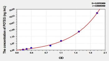
Human POTE Ankyrin Domain Family, Member G (POTEG) ELISA Kit [orb780419]
Human
0.32-20 ng/mL
0.121 ng/mL
48 T, 96 T - Item 1 of 4
- Item 1 of 4
- Item 1 of 4
- Item 1 of 4


Quality Guarantee
Explore bioreagents carefree to elevate your research. All our products are rigorously tested for performance. If a product does not perform as described on its datasheet, our scientific support team will provide expert troubleshooting, a prompt replacement, or a refund. For full details, please see our Terms & Conditions and Buying Guide. Contact us at [email protected].
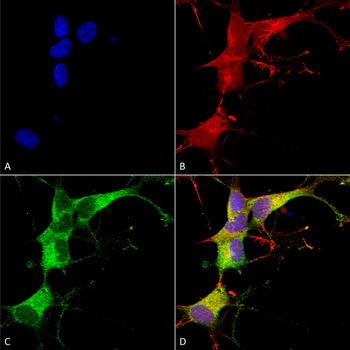
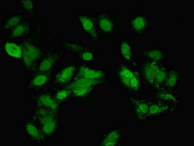
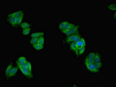
Immunocytochemistry/Immunofluorescence analysis using Mouse Anti-Ankyrin G Monoclonal Antibody, Clone N106/20. Tissue: Neuroblastoma cells (SH-SY5Y). Species: Human. Fixation: 4% PFA for 15 min. Primary Antibody: Mouse Anti-Ankyrin G Monoclonal Antibody at 1:100 for overnight at 4°C with slow rocking. Secondary Antibody: AlexaFluor 488 at 1:1000 for 1 hour at RT. Counterstain: Phalloidin-iFluor 647 (red) F-Actin stain; Hoechst (blue) nuclear stain at 1:800, 1.6mM for 20 min at RT. (A) Hoechst (blue) nuclear stain. (B) Phalloidin-iFluor 647 (red) F-Actin stain. (C) Ankyrin G Antibody (D) Composite.
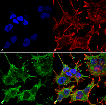
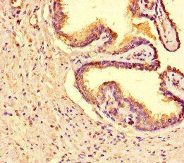
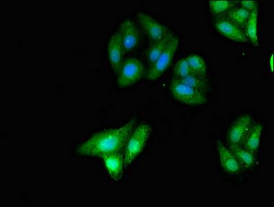
Immunocytochemistry/Immunofluorescence analysis using Mouse Anti-Ankyrin G Monoclonal Antibody, Clone N106/20. Tissue: Neuroblastoma cell line (SK-N-BE). Species: Human. Fixation: 4% Formaldehyde for 15 min at RT. Primary Antibody: Mouse Anti-Ankyrin G Monoclonal Antibody at 1:100 for 60 min at RT. Secondary Antibody: Goat Anti-Mouse ATTO 488 at 1:200 for 60 min at RT. Counterstain: Phalloidin Texas Red F-Actin stain; DAPI (blue) nuclear stain at 1:1000, 1:5000 for 60 min at RT, 5 min at RT. Localization: Cytoplasm. Magnification: 60X. (A) DAPI (blue) nuclear stain. (B) Phalloidin Texas Red F-Actin stain. (C) Ankyrin G Antibody. (D) Composite.
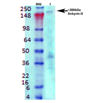
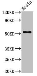
Western Blot analysis of Rat brain membrane lysate showing detection of Ankyrin G protein using Mouse Anti-Ankyrin G Monoclonal Antibody, Clone N106/20. Primary Antibody: Mouse Anti-Ankyrin G Monoclonal Antibody at 1:1000.
Quick Database Links
UniProt Details
− No UniProt data available
NCBI Gene Details
− No NCBI Gene data available
NCBI Reference Sequences
−Associated Accession Numbers
Curated reference sequences for the gene transcript and protein product| Protein | NP_066267.2 |
|---|
Documents Download
Datasheet
Product Information
Request a Document
Protocol Information
WB
Western Blot (IB, immunoblot)
IF
Immunofluorescence
ICC
Immunocytochemistry
Ankyrin G Antibody (orb67492)

- 0.0
Based on 0 reviews
Participating in our Biorbyt product reviews program enables you to support fellow scientists by sharing your firsthand experience with our products.
Login to Submit a ReviewAvailable Sizes
Select a size below
Choose Conjugation or Carrier Free Version
Free Secondary Antibody (20 ul)0/0
Please add an antibody product to your cart first.