You have no items in your shopping cart.
Description
Research Area
Cell Biology
Images & Validation
−Item 1 of 3
| Tested Applications | ELISA, IHC, WB |
|---|---|
| Dilution Range | ELISA: 1:20,000, IHC: 5-10 µg/mL, WB: 1:1,000 |
| Reactivity | Human |
| Application Notes |
Key Properties
−| Antibody Type | Primary Antibody |
|---|---|
| Host | Rat |
| Clonality | Monoclonal |
| Isotype | IgG2a |
| Clone No. | 25F2.D9 |
| Immunogen | This Protein G purified monoclonal antibody was produced in rats by repeated immunizations with full length recombinant mouse AHA1 protein followed by hybridoma development. |
| Target | Ahsa1 |
| Purity | This product is an IgG fraction antibody purified from tissue culture supernatant by Protein-G chromatography, followed by extensive dialysis against the buffer stated above. It is directed against human Aha1 protein. A BLAST analysis was used to suggest cross-reactivity with Aha1 protein from mouse, human and chimpanzee based on 100% homology with the immunizing sequence. Reactivity against homologues from other sources is not known. |
| Conjugation | Unconjugated |
Storage & Handling
−| Storage | Store vial at -20° C prior to opening. Aliquot contents and freeze at -20° C or below for extended storage. Avoid cycles of freezing and thawing. Centrifuge product if not completely clear after standing at room temperature. This product is stable for several weeks at 4° C as an undiluted liquid. Dilute only prior to immediate use. |
|---|---|
| Form/Appearance | Liquid (sterile filtered) |
| Buffer/Preservatives | Preservative: None. Stabilizer: None; Buffer: 0.02 M Potassium Phosphate, 0.15 M Sodium Chloride, pH 7.2 |
| Concentration | 1.0 mg/mL |
| Expiration Date | 12 months from date of receipt. |
| Dry Ice Shipping | Please note: This product requires shipment on dry ice. A dry ice surcharge will apply. |
| Disclaimer | For research use only |
Alternative Names
−rat anti-Aha1 Antibody, Aha1, Ahsa1 antibody, Activator of Hsp90 ATPase, Activator of 90 kDa heat shock protein ATPase homolog 1 antibody
Similar Products
−- Item 1 of 10
AHA1/AHSA1 Rabbit Polyclonal Antibody [orb1145840]
ELISA, FC, ICC, IF, IHC, WB
Human, Mouse, Rat
Rabbit
Polyclonal
Unconjugated
100 μg - Item 1 of 8
- Item 1 of 3
AHSA1 Rabbit Polyclonal Antibody [orb624569]
ELISA, IF, IHC, IP, WB
Human, Mouse, Rat
Rabbit
Polyclonal
Unconjugated
50 μg, 100 μg - Item 1 of 4
- Item 1 of 4
AHA1 Antibody (Biotin) [orb147670]
ELISA, ICC, IF, IHC, IP, WB
Human, Mouse, Rat
Rat
Monoclonal
Biotin
100 μg


Quality Guarantee
Explore bioreagents carefree to elevate your research. All our products are rigorously tested for performance. If a product does not perform as described on its datasheet, our scientific support team will provide expert troubleshooting, a prompt replacement, or a refund. For full details, please see our Terms & Conditions and Buying Guide. Contact us at [email protected].
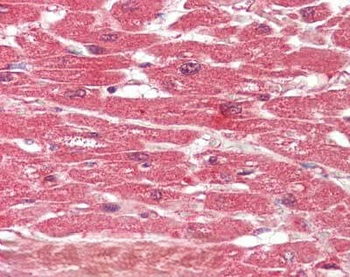
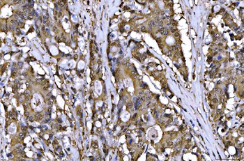
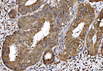
Immunohistochemistry of Rat anti-AHA1 antibody. Tissue: human heart. Fixation: formalin fixed paraffin embedded. Antigen retrieval: not required. Primary antibody: anti-AHA1 antibody at 10 µg/ml for 1 h at RT. Secondary antibody: Peroxidase rat secondary antibody at 1:10000 for 45 min at RT. Staining: AHA-1 as precipitated red signal with hematoxylin purple nuclear counterstain.
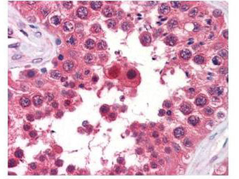
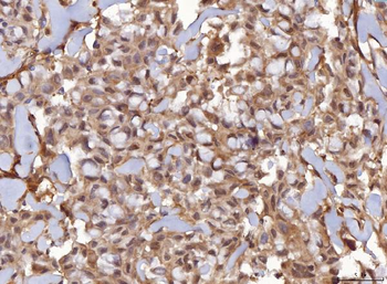
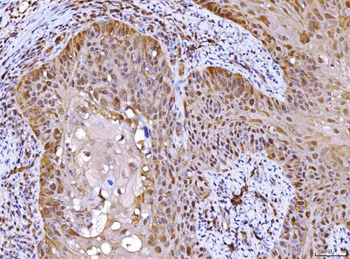
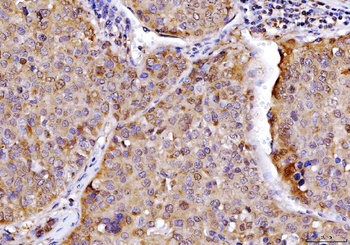
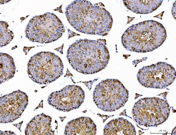
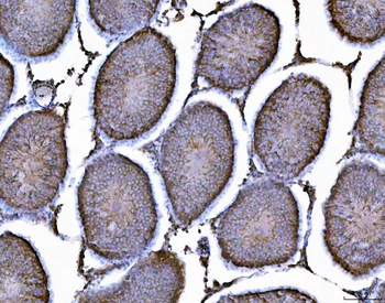
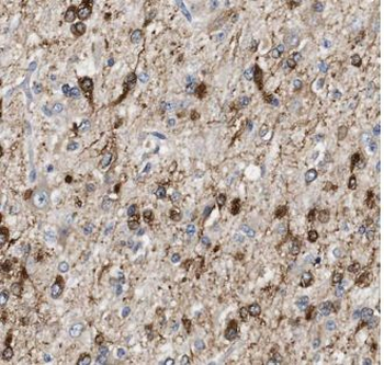
Biorbyt's anti-AHA1 monoclonal antibody was used at a 5-10 µg/ml to detect AHA1 in the seminiferous tubule of human testis (40X) showing moderate staining. Leydig cells showed faint to moderate staining. Expression of AHA1 is reported in many epithelial and lymphatic tissues, with cytoplasmic localization. This antibody showed moderate cytoplasmic staining of a variety of epithelial tissues and lymphoid organs such as spleen and tonsil with minimal background staining. The image shows the localization of the antibody as the precipitated red signal, with a hematoxylin purple nuclear counterstain. Tissue was formalin-fixed and paraffin embedded.
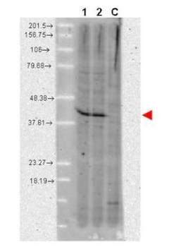
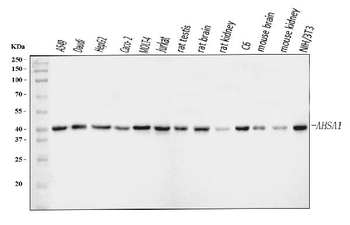
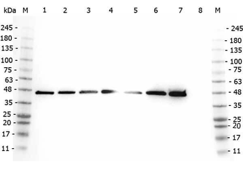
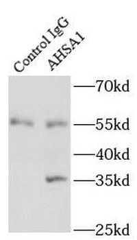
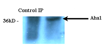
Western blot using Biorbyt's anti-AHA1 monoclonal antibody shows detection of a band ~42 kDa in size corresponding to AHA1. Molecular weight markers are shown at the left. Lane 1: A431 whole cell lysate (p/n orb348665) and Lane 2: MCF-7 whole cell lysate (p/n orb348664). A control lane is shown where primary antibody was omitted from the incubation (lane C). For best results, block the membrane overnight with 3% BSA in TBS followed by reaction with primary antibody diluted 1:1000 and use HRP conjugated anti-Rat IgG (p/n orb347790) secondary antibody diluted 1:20000 in blocking buffer (p/n orb348637) for detection.
Quick Database Links
UniProt Details
− No UniProt data available
NCBI Reference Sequences
−Associated Accession Numbers
Curated reference sequences for the gene transcript and protein product| Protein | NP_666148.1 |
|---|
Documents Download
Datasheet
Product Information
Request a Document
Protocol Information
WB
Western Blot (IB, immunoblot)
IHC
Immunohistochemistry
ELISA
Enzyme-linked Immunosorbent Assay (EIA)
Ahsa1 Antibody (orb345001)

- 0.0
Based on 0 reviews
Participating in our Biorbyt product reviews program enables you to support fellow scientists by sharing your firsthand experience with our products.
Login to Submit a ReviewAvailable Sizes
Select a size below
Choose Conjugation or Carrier Free Version
Free Secondary Antibody (20 ul)0/0
Please add an antibody product to your cart first.