You have no items in your shopping cart.
Description
Research Area
Pharmacology & Drug Discovery
Images & Validation
−Item 1 of 4
| Tested Applications | ELISA, ICC, IF, IP, WB |
|---|---|
| Dilution Range | WB (1:250), ICC/IF (1:100) |
| Reactivity | All |
| Application Notes |
Key Properties
−| Host | Rabbit |
|---|---|
| Clonality | Polyclonal |
| Immunogen | Acetylated KLH Conjugated |
| Target | Acetylated Lysine |
| Purification | Protein A Purified |
| Conjugation | Biotin |
Storage & Handling
−| Storage | Conjugated antibodies should be stored according to the product label |
|---|---|
| Buffer/Preservatives | 136.36mM Ethanolamine, 133.23 mM Chlorides, 9.55mM Phosphates, 9.55mM Sodium Bicarbonate. |
| Concentration | 0.25 mg/ml |
| Expiration Date | 12 months from date of receipt. |
| Disclaimer | For research use only |
Alternative Names
−N6-Acetyl-L-lysine, N-epsilon-Acetyl-L-lysine, Nepsilon-Acetyllysine, N6-Acetyllysine, Acetyllysine
Similar Products
−- Item 1 of 3
HSP70 (Acetyl Lys77) Antibody (Biotin) [orb414154]
ELISA, ICC, IF, IHC, WB
Human, Mouse, Rat
Rabbit
Polyclonal
Biotin
100 μl - Item 1 of 2
Acetylated Lysine Antibody (Biotin) [orb147381]
ELISA, ICC, IF, IHC, IP, WB
All
Mouse
Monoclonal
Biotin
100 μg - Item 1 of 1

Acetyl Lysine Rabbit Polyclonal Antibody (Biotin) [orb451632]
ELISA, ICC, IF, IHC-Fr, IHC-P
All
Rabbit
Polyclonal
Biotin
100 μlAcetyl Lysine Mouse Monoclonal Antibody (Biotin) [orb452997]
FC, ICC, IF, IHC-Fr, IHC-P
Human
All
Mouse
Monoclonal
Biotin
100 μl
Quality Guarantee
Explore bioreagents carefree to elevate your research. All our products are rigorously tested for performance. If a product does not perform as described on its datasheet, our scientific support team will provide expert troubleshooting, a prompt replacement, or a refund. For full details, please see our Terms & Conditions and Buying Guide. Contact us at [email protected].

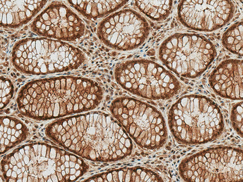
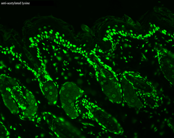
Immunocytochemistry/Immunofluorescence analysis using Rabbit Anti-Acetylated Lysine Polyclonal Antibody. Tissue: Heat Shocked Cervical cancer cell line (HeLa). Species: Human. Fixation: 2% Formaldehyde for 20 min at RT. Primary Antibody: Rabbit Anti-Acetylated Lysine Polyclonal Antibody at 1:100 for 12 hours at 4°C. Secondary Antibody: R-PE Goat Anti-Rabbit (yellow) at 1:200 for 2 hours at RT. Counterstain: DAPI (blue) nuclear stain at 1:40000 for 2 hours at RT. Localization: Nucleus. Cytoplasm. Magnification: 100x. (A) DAPI (blue) nuclear stain. (B) Anti-Acetylated Lysine Antibody. (C) Composite. Heat Shocked at 42°C for 1h.
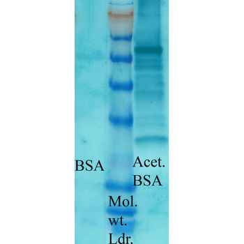
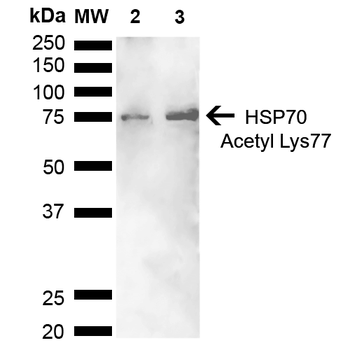
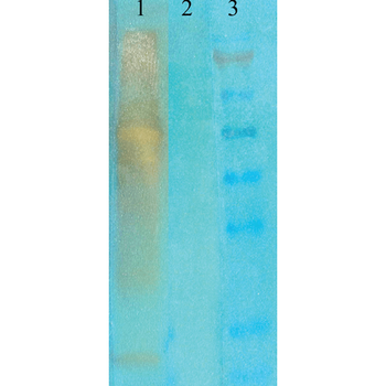
Western blot analysis of Bovine serum albumin showing detection of Acetylated Lysine protein using Rabbit Anti-Acetylated Lysine Polyclonal Antibody. Primary Antibody: Rabbit Anti-Acetylated Lysine Polyclonal Antibody at 1:1000. Acetylated lysine in BSA (Left) and Acetylated BSA (Right).
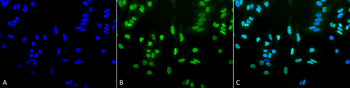
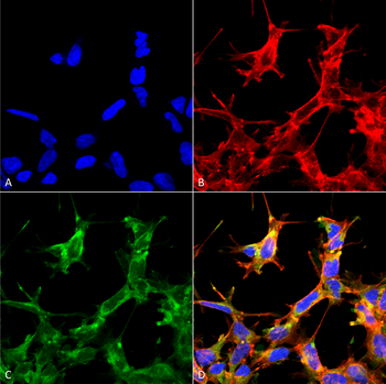
Immunocytochemistry/Immunofluorescence analysis using Rabbit Anti-Acetylated Lysine Polyclonal Antibody. Tissue: Heat Shocked Cervical cancer cell line (HeLa). Species: Human. Fixation: 2% Formaldehyde for 20 min at RT. Primary Antibody: Rabbit Anti-Acetylated Lysine Polyclonal Antibody at 1:100 for 12 hours at 4°C. Secondary Antibody: FITC Goat Anti-Rabbit (green) at 1:200 for 2 hours at RT. Counterstain: DAPI (blue) nuclear stain at 1:40000 for 2 hours at RT. Localization: Nucleus. Cytoplasm. Magnification: 20x. (A) DAPI (blue) nuclear stain. (B) Anti-Acetylated Lysine Antibody. (C) Composite. Heat Shocked at 42°C for 1h.
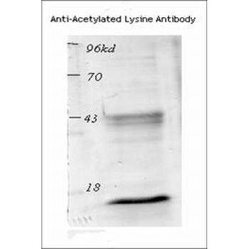
Western blot analysis of Mouse Spleen lysates showing detection of Acetylated Lysine protein using Rabbit Anti-Acetylated Lysine Polyclonal Antibody. Primary Antibody: Rabbit Anti-Acetylated Lysine Polyclonal Antibody at 1:1000.
Quick Database Links
Gene Symbol
Acetylated Lysine
Documents Download
Datasheet
Product Information
Request a Document
Protocol Information
WB
Western Blot (IB, immunoblot)
IF
Immunofluorescence
ICC
Immunocytochemistry
ELISA
Enzyme-linked Immunosorbent Assay (EIA)
IP
Immunoprecipitation
Acetylated Lysine Antibody (Biotin) (orb67532)

- 0.0
Based on 0 reviews
Participating in our Biorbyt product reviews program enables you to support fellow scientists by sharing your firsthand experience with our products.
Login to Submit a ReviewAvailable Sizes
Select a size below
Free Secondary Antibody (20 ul)0/0
Please add an antibody product to your cart first.