You have no items in your shopping cart.
YY1 Antibody
SKU: orb1927299
Description
Research Area
Epigenetics & Chromatin
Images & Validation
−Item 1 of 5
| Tested Applications | IHC-P, WB |
|---|---|
| Dilution Range | IHC-P - 1:100-500, WB - 1:1000 |
| Reactivity | Human, Monkey, Rat, Zebrafish |
Key Properties
−| Host | Mouse |
|---|---|
| Clonality | Monoclonal |
| Isotype | IgG2a |
| Molecular Weight | 44713 Da |
| Conjugation | Unconjugated |
Storage & Handling
−| Storage | Maintain refrigerated at 2-8°C for up to 2 weeks. For long term storage store at -20°C in small aliquots to prevent freeze-thaw cycles |
|---|---|
| Form/Appearance | Purified monoclonal antibody supplied in PBS with 0.09% (W/V) sodium azide. This antibody is purified through a protein G column, followed by dialysis against PBS. |
| Expiration Date | 12 months from date of receipt. |
| Disclaimer | For research use only |
Alternative Names
−INO80S
Similar Products
−- Item 1 of 18
YY1 Recombinant Rabbit Monoclonal Antibody (Nuclear Loading Control) [orb704397]
FC, IF, IHC-Fr, IHC-P, WB
Mouse, Rat
Human, Mouse, Rat
Rabbit
Recombinant
Unconjugated
50 μl, 100 μl - Item 1 of 17
HDAC2 Recombinant Rabbit Monoclonal Antibody [orb559204]
FC, ICC, IF, IHC-Fr, IHC-P, WB
Mouse, Rat
Human, Mouse, Rat
Rabbit
Recombinant
Unconjugated
50 μl, 100 μl - Item 1 of 10
HDAC2 Rabbit Polyclonal Antibody [orb13469]
IF, IHC-Fr, IHC-P
Bovine, Equine, Gallus, Porcine
Human, Mouse, Rat
Rabbit
Polyclonal
Unconjugated
50 μl, 100 μl, 200 μl - Item 1 of 9
YY1 Mouse Monoclonal Antibody [orb1145860]
FC, ICC, IF, IHC, WB
Human, Mouse, Rat
Mouse
Monoclonal
Unconjugated
100 μg - Item 1 of 6
YY1 Mouse Monoclonal Antibody [orb1145861]
FC, IHC, WB
Human, Mouse, Rat
Mouse
Monoclonal
Unconjugated
100 μg


Quality Guarantee
Explore bioreagents carefree to elevate your research. All our products are rigorously tested for performance. If a product does not perform as described on its datasheet, our scientific support team will provide expert troubleshooting, a prompt replacement, or a refund. For full details, please see our Terms & Conditions and Buying Guide. Contact us at [email protected].
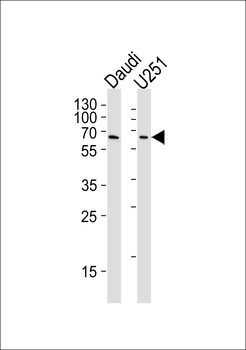
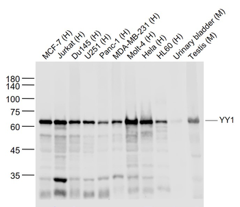
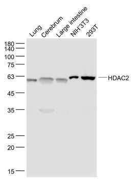
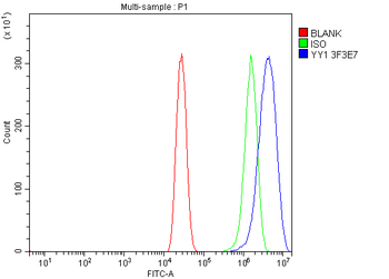
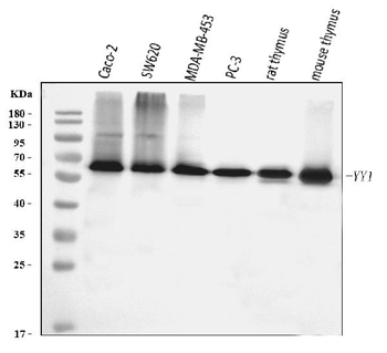
YY1 Antibody western blot analysis in Daudi and U251 cell line lysates (35μg/lane).This demonstrates the YY1 antibody detected the YY1 protein (arrow).
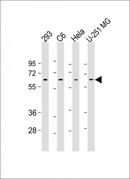
All lanes: Anti-YY1 Antibody (Center) at 1:500-1:1000 dilution. Lane 1: 293 whole cell lysate. Lane 2: C6 whole cell lysate. Lane 3: Hela whole cell lysate. Lane 4: U-251 MG whole cell lysate. Lysates/proteins at 20 µg per lane. Secondary Goat Anti-mouse IgG, (H+L), Peroxidase conjugated at 1/10000 dilution. Predicted band size: 45 kDa. Blocking/Dilution buffer: 5% NFDM/TBST.
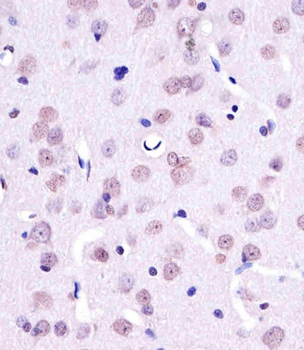
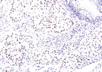
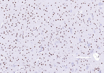
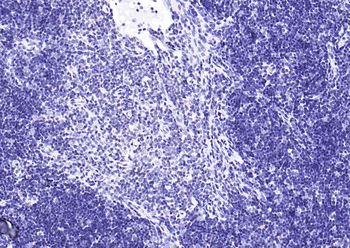
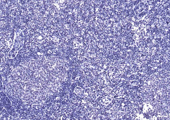
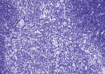
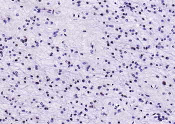
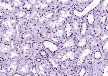
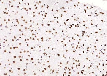
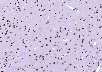
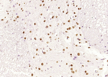
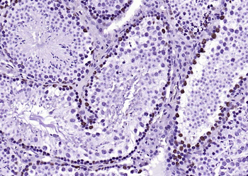
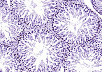
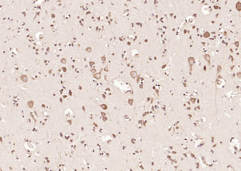
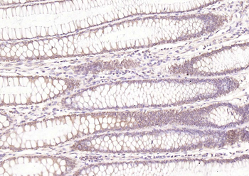
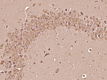
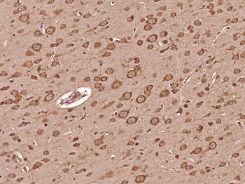
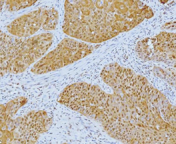
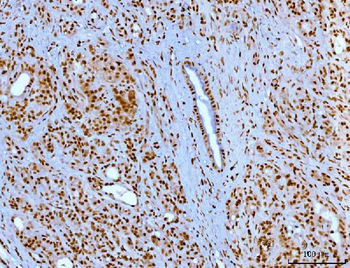
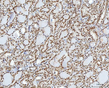
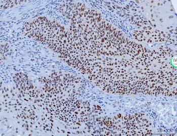
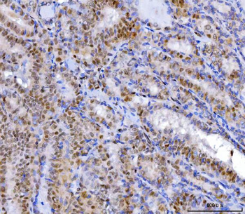
Staining YY1 in Rat brain tissue sections by Immunohistochemistry (IHC-P - paraformaldehyde-fixed, paraffin-embedded sections). Tissue was fixed with formaldehyde and blocked with 3% BSA for 0.5 hour at room temperature; antigen retrieval was by heat mediation with a citrate buffer (pH6). Samples were incubated with primary antibody (1/25) for 1 hours at 37°C. A undiluted biotinylated goat polyvalent antibody was used as the secondary antibody.

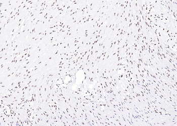
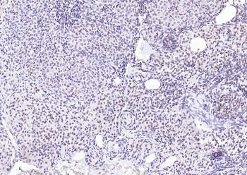
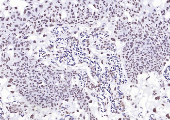
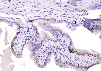
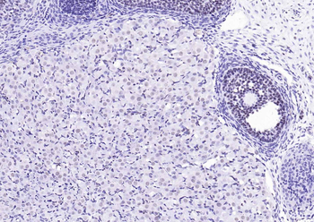
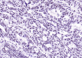
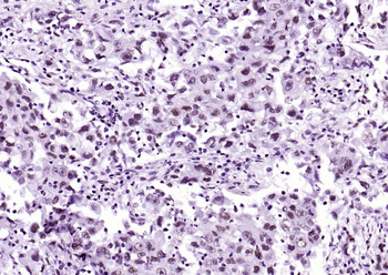
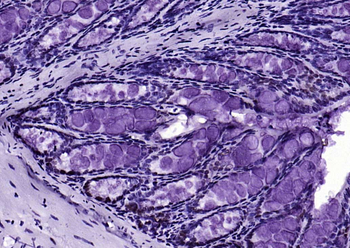
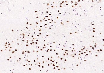
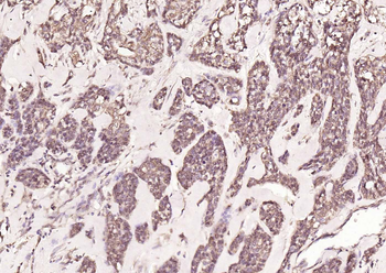
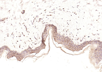
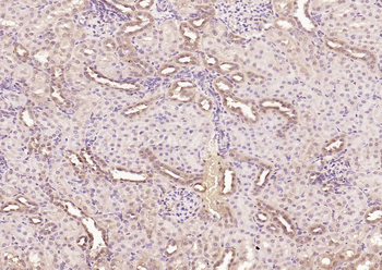
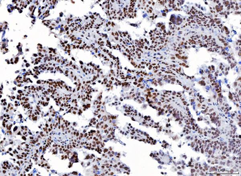
Staining YY1 in Monkey brain tissue sections by Immunohistochemistry (IHC-P - paraformaldehyde-fixed, paraffin-embedded sections). Tissue was fixed with formaldehyde and blocked with 3% BSA for 0.5 hour at room temperature; antigen retrieval was by heat mediation with a citrate buffer (pH6). Samples were incubated with primary antibody (1/25) for 1 hours at 37°C. A undiluted biotinylated goat polyvalent antibody was used as the secondary antibody.
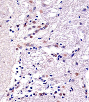
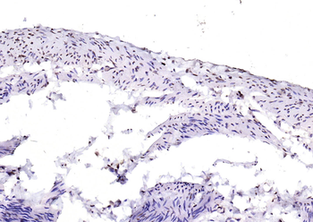
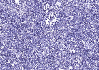
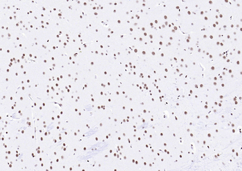
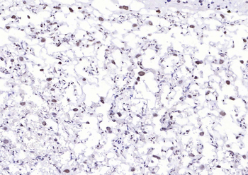
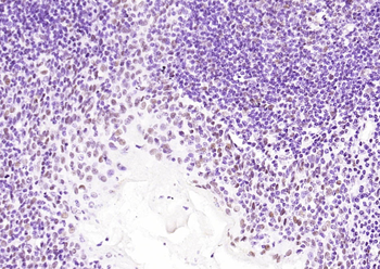
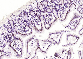
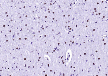
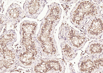
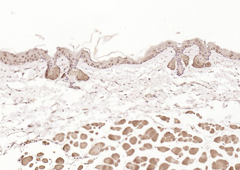
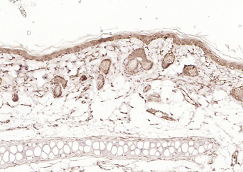
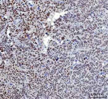
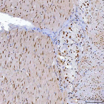
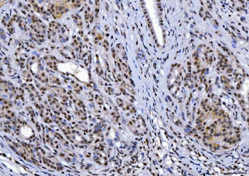
Staining YY1 in Zebra fish brain tissue sections by Immunohistochemistry (IHC-P - paraformaldehyde-fixed, paraffin-embedded sections). Tissue was fixed with formaldehyde and blocked with 3% BSA for 0.5 hour at room temperature; antigen retrieval was by heat mediation with a citrate buffer (pH6). Samples were incubated with primary antibody (1/25) for 1 hours at 37°C. A undiluted biotinylated goat polyvalent antibody was used as the secondary antibody.
Quick Database Links
UniProt
UniProt Details
− No UniProt data available
Documents Download
Datasheet
Product Information
Request a Document
Protocol Information
WB
Western Blot (IB, immunoblot)
IHC-P
Immunohistochemistry Paraffin
YY1 Antibody (orb1927299)

- 0.0
Based on 0 reviews
Participating in our Biorbyt product reviews program enables you to support fellow scientists by sharing your firsthand experience with our products.
Login to Submit a ReviewAvailable Sizes
Select a size below