You have no items in your shopping cart.
TrkA-pY791 Antibody
SKU: orb1928982
Description
Research Area
Signal Transduction
Images & Validation
−Item 1 of 6
| Tested Applications | FC, IHC-P, WB |
|---|---|
| Dilution Range | WB: 1:1000, WB: 1:2000, IHC-P: 1:10~50, IHC-P: 1:25, FC: 1:10~50, FC: 1:25 |
| Reactivity | Human, Mouse |
Key Properties
−| Antibody Type | Primary Antibody |
|---|---|
| Host | Rabbit |
| Clonality | Polyclonal |
| Isotype | Rabbit IgG |
| Clone No. | RB16572 |
| Target | This TrkA antibody is generated from rabbits immunized with a KLH conjugated synthetic peptide between 769-796 amino acids from human TrkA. |
| Molecular Weight | 87497 Da |
| Conjugation | Unconjugated |
Storage & Handling
−| Storage | Maintain refrigerated at 2-8°C for up to 2 weeks. For long term storage store at -20°C in small aliquots to prevent freeze-thaw cycles |
|---|---|
| Form/Appearance | Purified polyclonal antibody supplied in PBS with 0.09% (W/V) sodium azide. This antibody is purified through a protein A column, followed by peptide affinity purification. |
| Expiration Date | 12 months from date of receipt. |
| Disclaimer | For research use only |
Alternative Names
−High affinity nerve growth factor receptor, Neurotrophic tyrosine kinase receptor type 1, TRK1-transforming tyrosine kinase protein, Tropomyosin-related kinase A, Tyrosine kinase receptor, Tyrosine kinase receptor A, Trk-A, gp140trk, p140-TrkA, NTRK1, MTC, TRK, TRKA
Similar Products
−
TrkA-pY791 Antibody [orb2996479]
FC, IHC-P, WB
Human, Mouse, Rat
Rabbit
Polyclonal
Unconjugated
100 μl, 50 μl
Quality Guarantee
Explore bioreagents carefree to elevate your research. All our products are rigorously tested for performance. If a product does not perform as described on its datasheet, our scientific support team will provide expert troubleshooting, a prompt replacement, or a refund. For full details, please see our Terms & Conditions and Buying Guide. Contact us at [email protected].
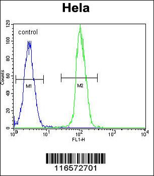
TrkA-pY791 Antibody flow cytometric analysis of Hela cells (right histogram) compared to a negative control cell (left histogram). FITC-conjugated goat-anti-rabbit secondary antibodies were used for the analysis.
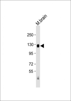
Anti-TrkA (Y791) Antibody at 1:2000 dilution + mouse brain lysate. Lysates/proteins at 20 µg per lane. Secondary Goat Anti-Rabbit IgG, (H+L), Peroxidase conjugated at 1/10000 dilution. Predicted band size: 87 kDa. Blocking/Dilution buffer: 5% NFDM/TBST.
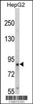
Western blot analysis of hTrkA-pY791 in HepG2 cell line lysates (35 ug/lane). TRK (arrow) was detected using the purified Pab.
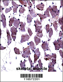
TrkA-pY791 Antibody immunohistochemistry analysis in formalin fixed and paraffin embedded human skeletal muscle followed by peroxidase conjugation of the secondary antibody and DAB staining.This data demonstrates the use of TrkA-pY791 Antibody for immunohistochemistry. Clinical relevance has not been evaluated.
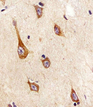
Staining TrkA in human brain tissue sections by Immunohistochemistry (IHC-P - paraformaldehyde-fixed, paraffin-embedded sections). Tissue was fixed with formaldehyde and blocked with 3% BSA for 0.5 hour at room temperature; antigen retrieval was by heat mediation with a citrate buffer (pH6). Samples were incubated with primary antibody (1/25) for 1 hours at 37°C. A undiluted biotinylated goat polyvalent antibody was used as the secondary antibody.
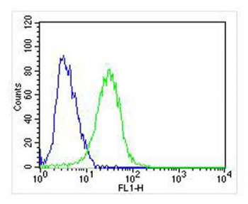
Overlay histogram showing SH-SY5Y cells stained (green line). The cells were fixed with 2% paraformaldehyde (10 min). The cells were then icubated in 2% bovine serum albumin to block non-specific protein-protein interactions followed by the antibody (1:25 dilution) for 60 min at 37°C. The secondary antibody used was Goat-Anti-Rabbit IgG, DyLight 488 Conjugated Highly Cross-Adsorbed at 1/400 dilution for 40 min at 37°C. Isotype control antibody (blue line) was rabbit IgG (1 μg/1x10^6 cells) used under the same conditions. Acquisition of > 10000 events was performed.
Quick Database Links
Gene Symbol
This TrkA antibody is generated from rabbits immunized with a KLH conjugated synthetic peptide between 769-796 amino acids from human TrkA.
UniProt
RefSeq (Protein):NP_002520.2, NP_001007793.1, NP_001012331.1
UniProt Details
− No UniProt data available
NCBI Reference Sequences
−Associated Accession Numbers
Curated reference sequences for the gene transcript and protein product| Protein | NP_002520.2, NP_001007793.1, NP_001012331.1 |
|---|
Documents Download
Datasheet
Product Information
Request a Document
Protocol Information
WB
Western Blot (IB, immunoblot)
IHC-P
Immunohistochemistry Paraffin
FC
Flow Cytometry
TrkA-pY791 Antibody (orb1928982)

- 0.0
Based on 0 reviews
Participating in our Biorbyt product reviews program enables you to support fellow scientists by sharing your firsthand experience with our products.
Login to Submit a ReviewAvailable Sizes
Select a size below