You have no items in your shopping cart.
Description
Research Area
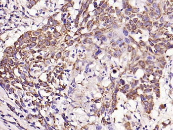
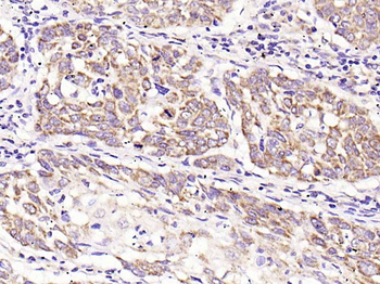
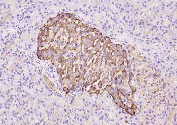
Cancer Biology
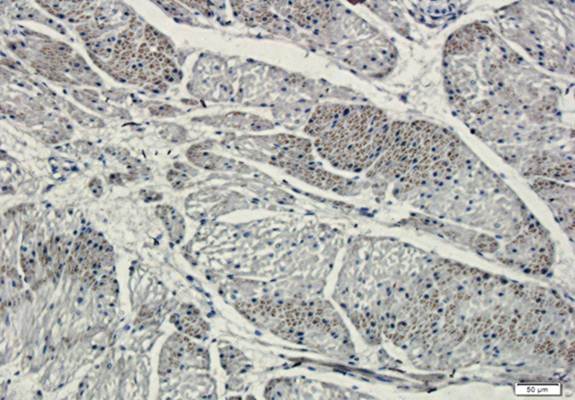
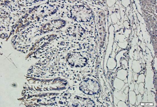
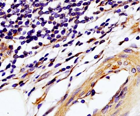
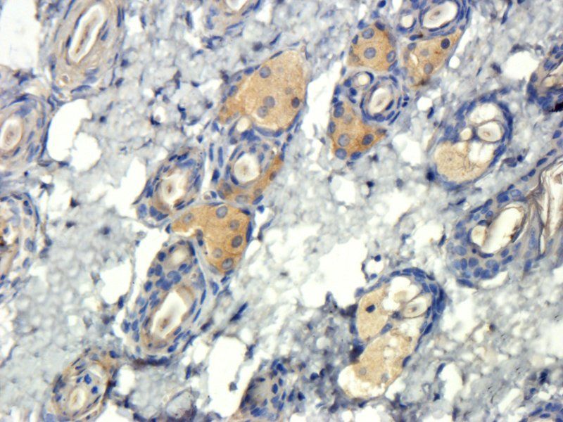
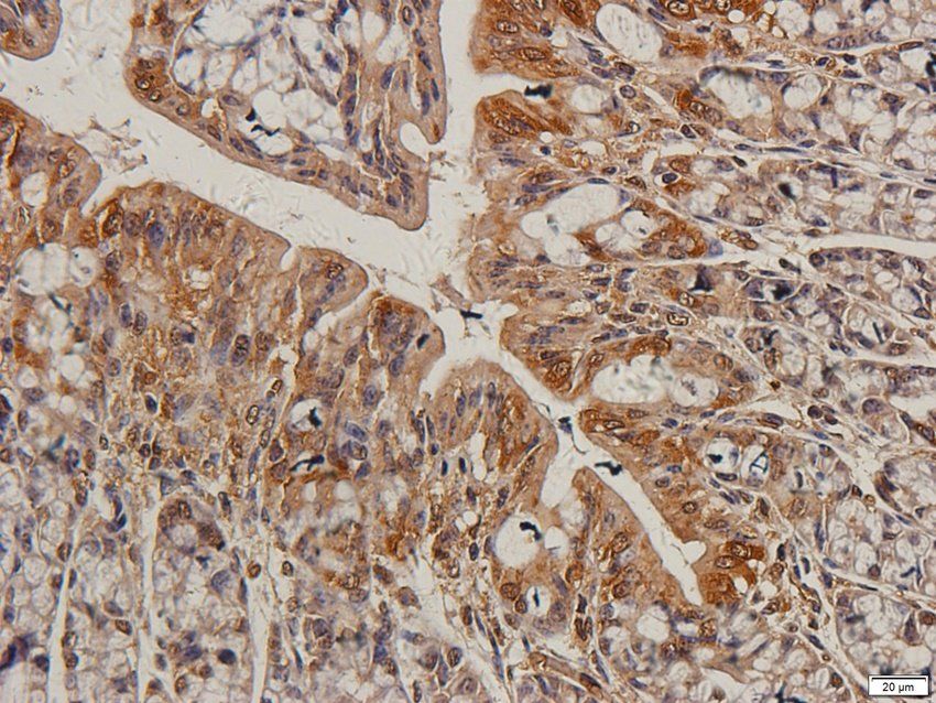
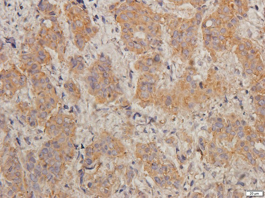
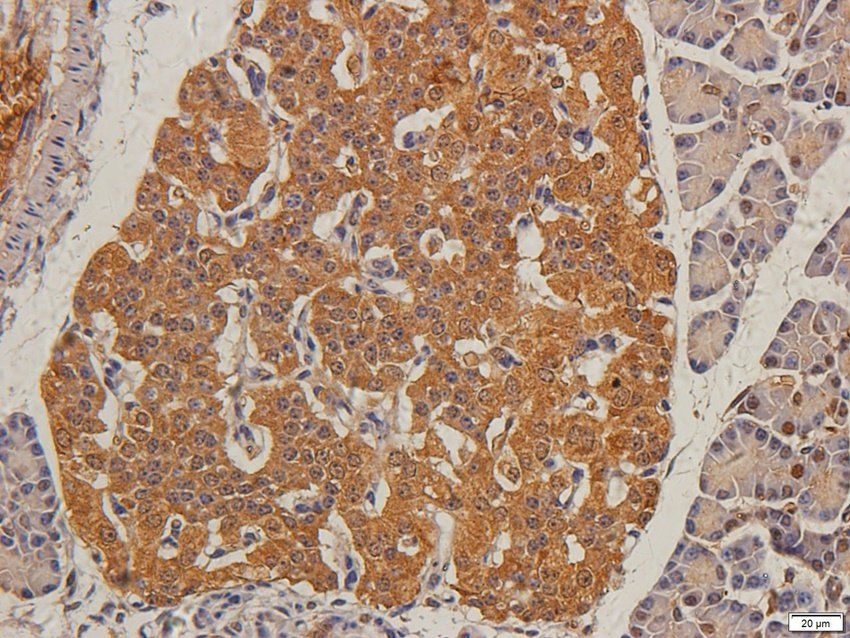
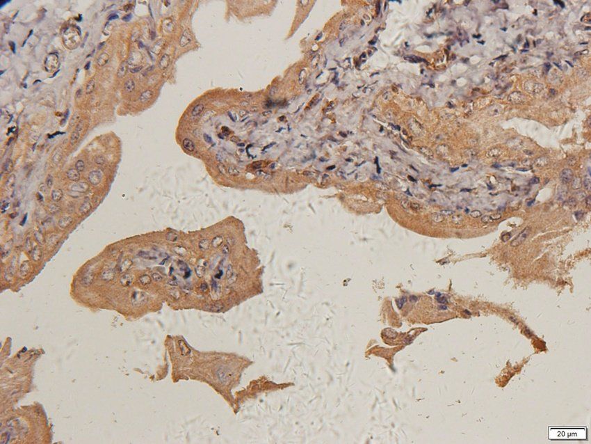
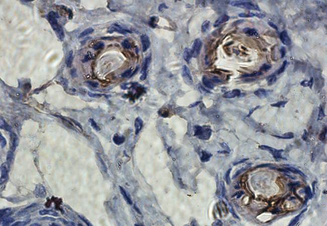
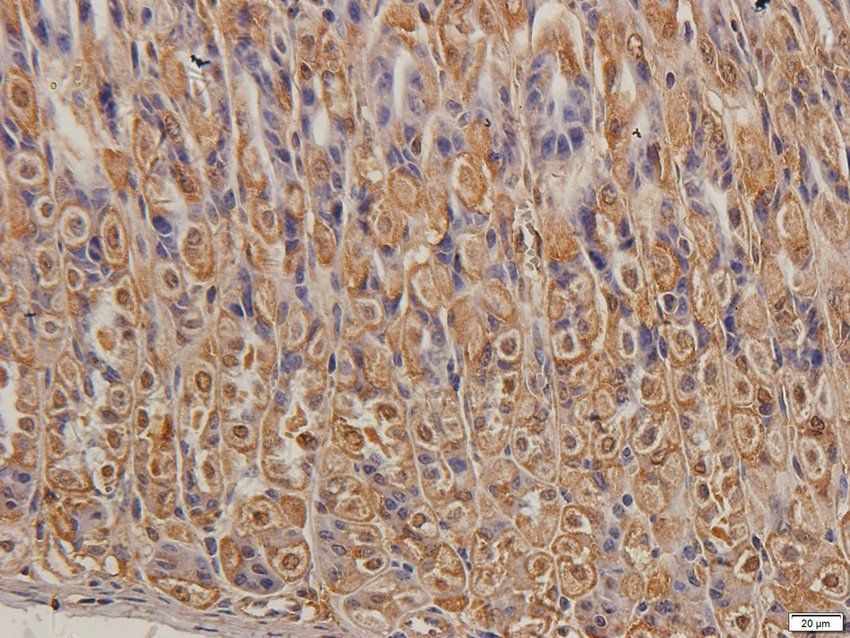
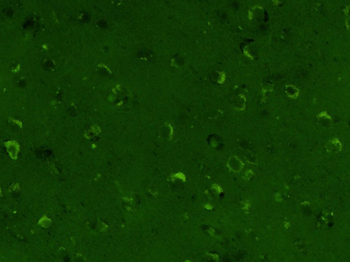
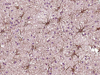
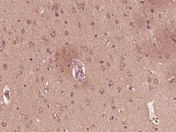
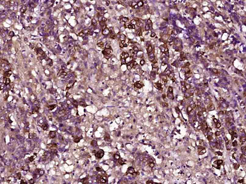
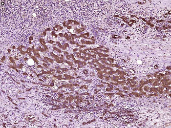
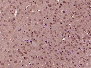
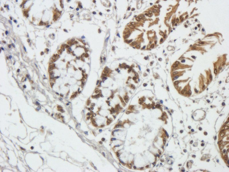
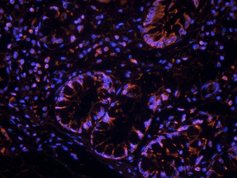
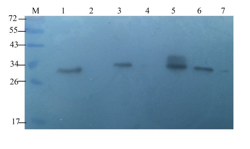
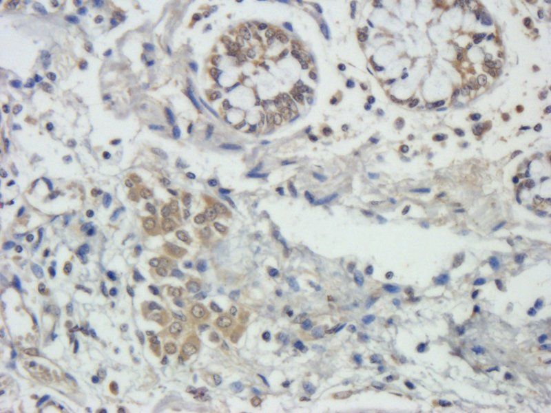
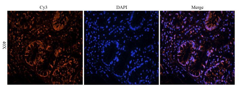
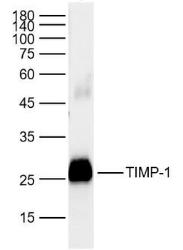
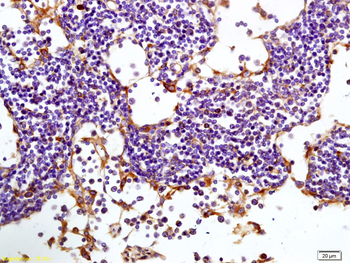
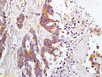
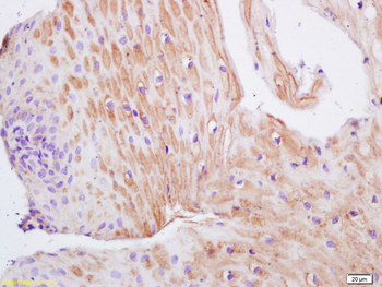
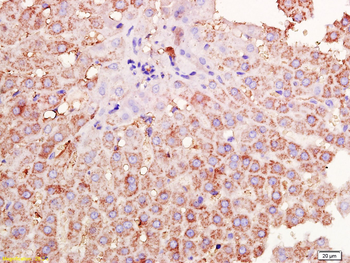
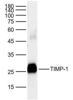
Images & Validation
−| Application Notes |
|---|
Key Properties
−| Source | Sf9, Baculovirus cells |
|---|---|
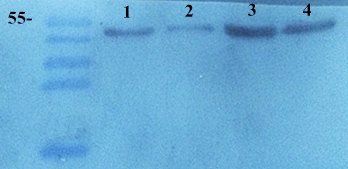
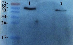
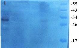
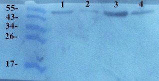
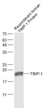
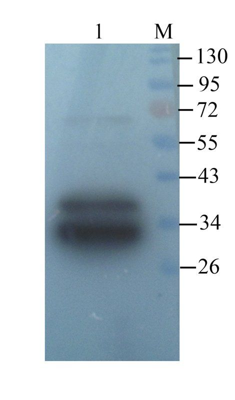
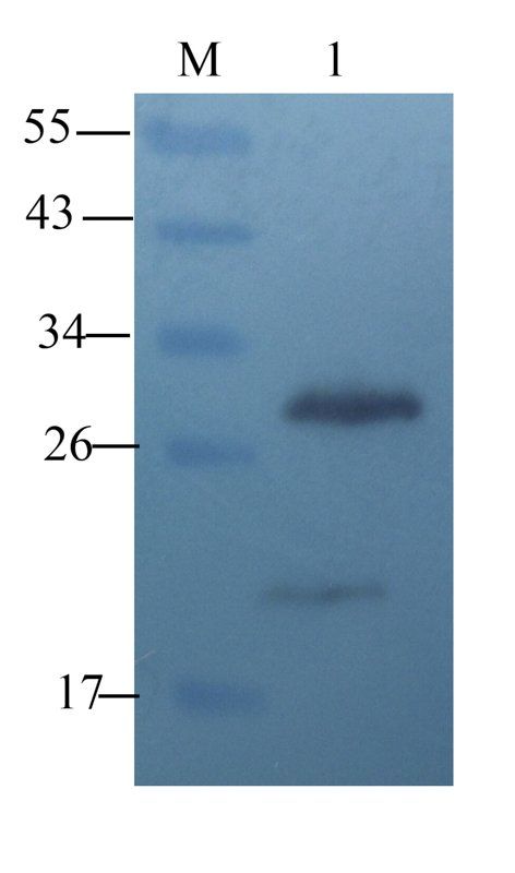
| Purity | Greater than 90.0% as determined by SDS-PAGE. |
| Protein Sequence | CSCAPTHPQT AFCNSDLVIR AKFMGSPEII ETTLYQRYEI KMTKMLKGFD AVGNATGFRF AYTPAMESLC GYVHKSQNRS EEFLIAGRLR NGNLHITACS FLVPWHNLSP AQQKAFVKTY SAGCGVCTVF PCSAIPCKLE SDSHCLWTDQ ILMGSEKGYQ SDHFACLPRN PDLCTWQYLG VSMTRSLPLA KAEAHHHHHH |
Storage & Handling
−| Storage | Stability: Store at 4°C if entire vial will be used within 2-4 weeks. Store, frozen at -20°C for longer periods of time. For long term storage it is recommended to add a carrier protein (0.1% HSA or BSA).Avoid multiple freeze-thaw cycles |
|---|---|
| Form/Appearance | Sterile filtered colorless solution. |
| Buffer/Preservatives | TIMP1 Ligand protein solution (0.5mg/ml) contains Phosphate buffered saline (pH7.4) and 10% glycerol. |
| Expiration Date | 6 months from date of receipt. |
| Disclaimer | For research use only |
Alternative Names
−Metalloproteinase inhibitor 1, Tissue inhibitor of metalloproteinases 1, TIMP-1, TIMP1.
Similar Products
−- Item 1 of 18
TIMP1 Rabbit Polyclonal Antibody [orb195994]
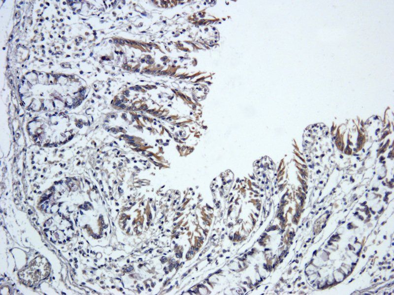
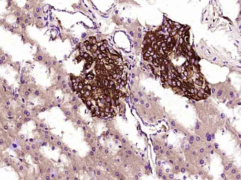
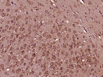
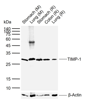
ELISA, ICC, IF, IHC-P, WB
Bovine, Canine, Guinea pig, Human, Mouse, Porcine, Rat, Sheep
Rabbit
Polyclonal
Unconjugated
100 μg - Item 1 of 10
TIMP-1 Mouse Monoclonal Antibody [orb500828]
FC, ICC
Mouse, Rat
Human
Mouse
Monoclonal
Unconjugated
100 μl, 50 μl, 200 μl, 200 μg - Item 1 of 7
TIMP1 Rabbit Polyclonal Antibody [orb11483]
ELISA, ICC, IF, IHC-P, WB
Rabbit
Polyclonal
Unconjugated
100 μg - Item 1 of 5
TIMP-1 Rabbit Polyclonal Antibody [orb313247]
WB
Bovine, Canine, Porcine, Rat, Sheep
Human
Rabbit
Polyclonal
Unconjugated
100 μl, 200 μl, 50 μl - Item 1 of 5
TIMP-1 Rabbit Polyclonal Antibody [orb100174]
WB
Bovine, Canine, Mouse, Porcine, Rabbit, Sheep
Human
Rabbit
Polyclonal
Unconjugated
100 μl, 200 μl, 50 μl
Quality Guarantee
Explore bioreagents carefree to elevate your research. All our products are rigorously tested for performance. If a product does not perform as described on its datasheet, our scientific support team will provide expert troubleshooting, a prompt replacement, or a refund. For full details, please see our Terms & Conditions and Buying Guide. Contact us at [email protected].
Documents Download
Datasheet
Product Information
Request a Document
Protocol Information
Protein Handling and Storage Guide
Protein Handling Guide
TIMP1 Protein (orb428855)

- 0.0
Based on 0 reviews
Participating in our Biorbyt product reviews program enables you to support fellow scientists by sharing your firsthand experience with our products.
Login to Submit a ReviewAvailable Sizes
Select a size below