You have no items in your shopping cart.
Description
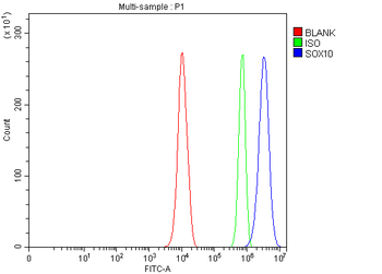
Research Area
Cancer Biology
Images & Validation
−Item 1 of 10
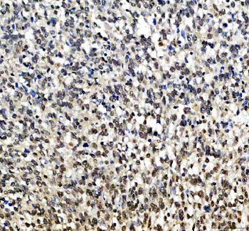
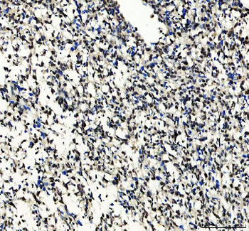
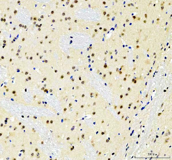
| Tested Applications | IHC, WB |
|---|---|
| Reactivity | Human |
| Predicted Reactivity | Bovine, Canine, Equine, Guinea pig, Mouse, Rabbit, Rat |
Key Properties
−| Host | Rabbit |
|---|---|
| Clonality | Polyclonal |
| Immunogen | The immunogen is a synthetic peptide directed towards the middle region of human SOX10 |
| Target | SOX10 |
| Protein Sequence | Synthetic peptide located within the following region: PGGEAEQGGTAAIQAHYKSAHLDHRHPGEGSPMSDGNPEHPSGQSHGPPT |
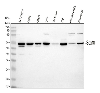
| Molecular Weight | 50 kDa |
| Purification | Affinity Purified |
| Conjugation | Unconjugated |
Storage & Handling
−| Storage | Maintain refrigerated at 2-8°C for up to 2 weeks. For long term storage store at -20°C in small aliquots to prevent freeze-thaw cycles. |
|---|---|
| Buffer/Preservatives | Liquid. Purified antibody supplied in 1x PBS buffer with 0.09% (w/v) sodium azide and 2% sucrose. |
| Concentration | 0.5 mg/ml |
| Expiration Date | 12 months from date of receipt. |
| Disclaimer | For research use only |
Alternative Names
−DOM, WS4, PCWH, WS2E, WS4C
Similar Products
−- Item 1 of 5
SOX10 Antibody (Center) [orb1930327]
IHC-P, WB
Porcine
Human, Mouse, Rat
Rabbit
Polyclonal
Unconjugated
50 μl, 100 μl - Item 1 of 3
SOX10 Rabbit Polyclonal Antibody [orb500740]
FC, WB
Bovine, Canine, Human, Porcine, Rabbit, Sheep
Mouse, Rat
Rabbit
Polyclonal
Unconjugated
50 μl, 100 μl, 200 μl - Item 1 of 4
SOX10 Rabbit Polyclonal Antibody [orb402212]
IHC, WB
Human, Mouse, Rat
Rabbit
Polyclonal
Unconjugated
100 μg - Item 1 of 3
SOX10 Rabbit Polyclonal Antibody [orb1743816]
ELISA, FC, ICC, IF, WB
Human, Mouse, Rat
Rabbit
Polyclonal
Unconjugated
100 μg - Item 1 of 3
SOX10 Rabbit Polyclonal Antibody [orb668233]
IF, IHC, WB
Human, Mouse, Rat
Rabbit
Polyclonal
Unconjugated
50 μl, 100 μl, 200 μl, 30 μl
Quality Guarantee
Explore bioreagents carefree to elevate your research. All our products are rigorously tested for performance. If a product does not perform as described on its datasheet, our scientific support team will provide expert troubleshooting, a prompt replacement, or a refund. For full details, please see our Terms & Conditions and Buying Guide. Contact us at [email protected].
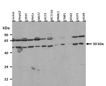
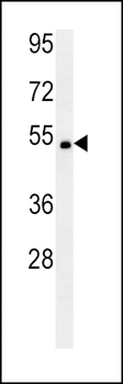
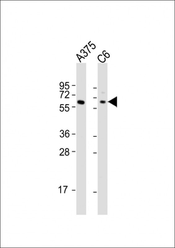
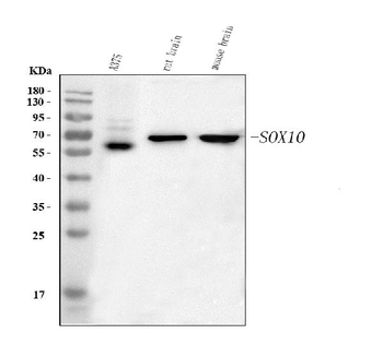
25 ug of the indicated Human whole cell extracts was loaded onto a 12% SDS-PAGE gel. 1 ug/ml of the antibody was used in this experiment.
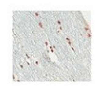
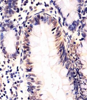
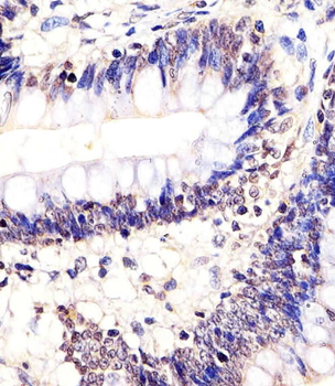
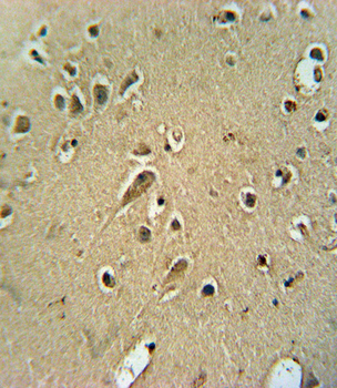
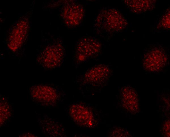
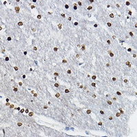
A/ [IHC-PZ] (optimal processing) human optic nerve (short post mortem interval) fixed by immersion in zinc-based fixative (BD Pharmingen 552658), processed to minimize antigen loss (shortened protocol, reduced exposure to high temperature); embedded in paraffin wax; sectioned at 3 microns A2/ [IHC-PZ + FF] As above for initial fixation then post-fixed in 10% buffered formalin for 5 days B/ [IHC-P] human optic nerve and spinal cord fixed in 10% buffered formalin (relatively short post mortem interval and fixation duration); standard processing; embedded in paraffin wax; sectioned at 3 microns Controls Negative: omission of primary. Positive: Olig2 reactivity was in comparison with antibody used at 1:10000 (IHC-PZ); 1:4000 (IHC-P).
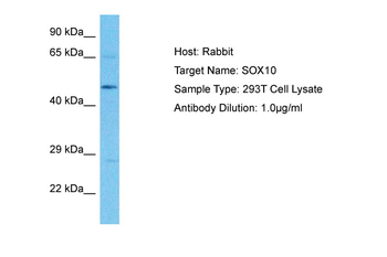
Sample Tissue: Human 293T Whole Cell, Antibody dilution: 3 ug/ml.

Sample Tissue: Human MCF7 Whole Cell, Antibody dilution: 1 ug/ml.
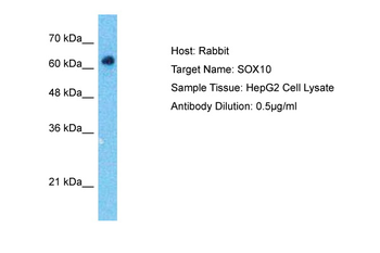
Sample Type: HepG2 Whole Cell lysates, Antibody dilution: 0.5 ug/ml.
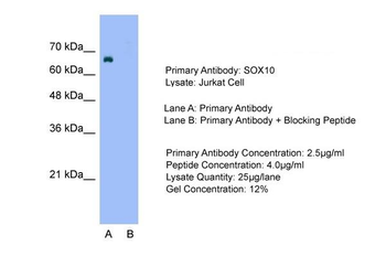
Sample Type: Jurkat cell lysates, Lane A: Primary Antibody, Lane B: Primary Antibody + Blocking Peptide, Primary Antibody Concentration: 2.5 ug/ml, Peptide Concentration: 4.0 ug/ml, Lysate Quantity: 25 ug/lane, Gel Concentration: 12%.
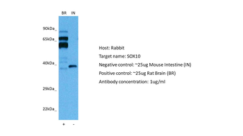
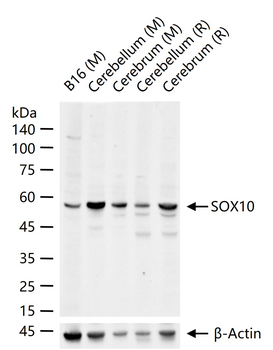
Positive control (+): Rat brain (R-BR), Negative control (-): Mouse intestine (M-IN), Antibody concentration: 1 ug/ml.
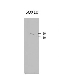
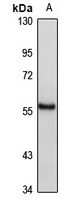
SOX10 antibody - middle region (orb574604) validated by WB using Hek 293 Whole Cell Lysate at 1:4, 000.
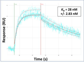
Surface Plasmon Resonance Kinetic Characterization of Polyclonal Antibody Affinity. Purified polyclonal antibodies were immobilized on a Protein A/G coated Carterra LSA sensor chip (PAGH200M) at concentrations of 5, and 50 ug/ml in duplicate. Antibodies on the surface were exposed to interaction with peptides sequentially via microfluidic controlled flow at 333nM peptide concentration for kinetic characterization of the binders for affinity and specificity, followed by curve fitting using the Kinetics software. Kd determinations for both concentrations were averaged and results and standard deviation are shown.
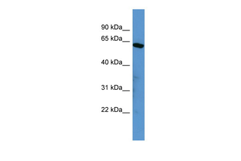
WB Suggested Anti-SOX10 Antibody Titration: 25 ug/ml, ELISA Titer: 1:62500, Positive Control: HepG2 cell lysate.
Documents Download
Datasheet
Product Information
Request a Document
Protocol Information
WB
Western Blot (IB, immunoblot)
IHC
Immunohistochemistry
SOX10 Rabbit Polyclonal Antibody (orb574604)

- 0.0
Based on 0 reviews
Participating in our Biorbyt product reviews program enables you to support fellow scientists by sharing your firsthand experience with our products.
Login to Submit a ReviewAvailable Sizes
Select a size below
Free Secondary Antibody (20 ul)0/0
Please add an antibody product to your cart first.