You have no items in your shopping cart.
Rhodopsin Antibody
SKU: orb1822458
Featured
Description
Research Area
Neuroscience, Signal Transduction
Images & Validation
−Item 1 of 3
| Tested Applications | ELISA, ICC, IF, IHC, IP, WB |
|---|---|
| Dilution Range | WB (1:1000), IHC (1000); optimal dilutions for assays should be determined by the user. |
| Reactivity | Amphibian, Aves, Bovine, Fish, Human, Mouse |
| Application Notes |
Key Properties
−| Host | Mouse |
|---|---|
| Clonality | Monoclonal |
| Isotype | IgG1 |
| Clone No. | 4D2 |
| Immunogen | Bovine Rhodopsin |
| Target | Rhodopsin |
| Molecular Weight | 40kDa |
| Purification | Protein G Purified |
| Conjugation | Unconjugated |
Storage & Handling
−| Storage | Maintain refrigerated at 2-8°C for up to 2 weeks. For long term storage store at -20°C in small aliquots to prevent freeze-thaw cycles. |
|---|---|
| Buffer/Preservatives | PBS pH 7.4, 50% glycerol, 0.09% sodium azide Storage buffer changes when conjugated |
| Concentration | 1 mg/ml |
| Expiration Date | 12 months from date of receipt. |
| Disclaimer | For research use only |
Alternative Names
−OPN2, opsd, opsin 2, opsin 2 rod pigment, opsin2, RHO, RP4, MGC138309, Retinitis Pigmentosa 4
Similar Products
−- Item 1 of 4
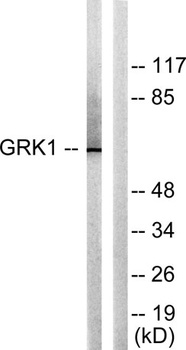
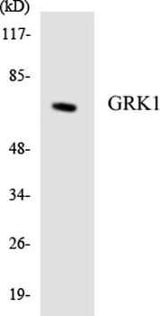
GRK 1 rabbit pAb Antibody [orb765351]
ELISA, IF, IHC, WB
Human, Mouse, Rat
Polyclonal
Unconjugated
50 μl, 100 μl - Item 1 of 1
- Item 1 of 1
- Item 1 of 1
- Item 1 of 3
Anti-Rhodopsin [Rho 1D4] [orb613825]
ELISA, IF, IHC, IP, WB
Amphibian, Bovine, Human, Mouse, Rat, Zebrafish
Mouse
Monoclonal
Unconjugated
0.2 mg
![Anti-Rhodopsin [Rho 1D4]](/images/pub/media/catalog/product/NewWebsite/35/orb613825_1.png)
![Anti-Rhodopsin [Rho 1D4]](/images/pub/media/catalog/product/NewWebsite/35/orb613825_2.png)
![Anti-Rhodopsin [Rho 1D4]](/images/pub/media/catalog/product/NewWebsite/35/orb613825_3.png)
Quality Guarantee
Explore bioreagents carefree to elevate your research. All our products are rigorously tested for performance. If a product does not perform as described on its datasheet, our scientific support team will provide expert troubleshooting, a prompt replacement, or a refund. For full details, please see our Terms & Conditions and Buying Guide. Contact us at [email protected].
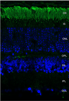
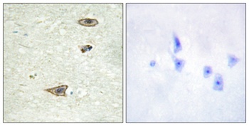
Immunohistochemistry analysis using Mouse Anti-Rhodopsin Monoclonal Antibody, Clone 4D2. Tissue: retina. Species: Mouse. Primary Antibody: Mouse Anti-Rhodopsin Monoclonal Antibody at 1:1000. Secondary Antibody: FITC Goat Anti-Mouse (green). Counterstain: DAPI (blue) nuclear stain. Localization: Staining of photoreceptor outer segment (OS). Other layers of the retina: IS – inner segment; ONL – outer nuclear layer; OPL – outer plexiform layer; INL – inner nuclear layer; IPL – inner plexiform layer; GCL – ganglion cell layer.
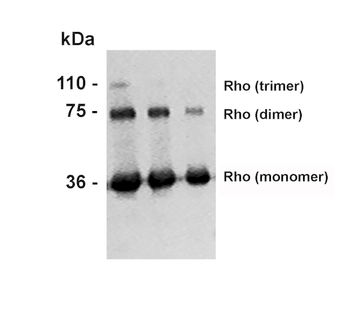
Western Blot analysis of Bovine photoreceptor membranes showing detection of Rhodopsin protein using Mouse Anti-Rhodopsin Monoclonal Antibody, Clone 4D2. Lane 1: MW ladder. Lane 2: 10 ug. Lane 3: 5 ug. Lane 4: 2.5 ug. Primary Antibody: Mouse Anti-Rhodopsin Monoclonal Antibody at 1:1000.
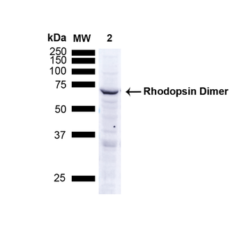
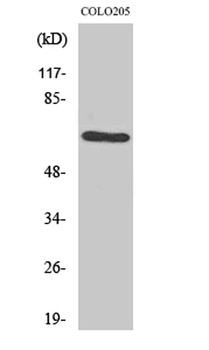
Western Blot analysis of Human A549 cells showing detection of ~38.9kDa Rhodopsin protein using Mouse Anti-Rhodopsin Monoclonal Antibody, Clone 4D2. Lane 1: MW ladder. Lane 2: Human A549 Cells 15 ug). Load: 15 ug. Block: 5% Skim Milk Powder in TBST. Primary Antibody: Mouse Anti-Rhodopsin Monoclonal Antibody at 1:1000 for 2.5 hours at RT with shaking. Secondary Antibody: Goat anti-mouse IgG:HRP at 1:1000 for 1 hour at RT with shaking. Color Development: Chemiluminescent for HRP (Moss) for 5 min in RT. Predicted/Observed Size: ~38.9kDa. Other Band (s): Band appears at ~75 kDa indicating detection of the Rhodopsin dimer.
Quick Database Links
UniProt Details
− No UniProt data available
NCBI Gene Details
− No NCBI Gene data available
NCBI Reference Sequences
−Associated Accession Numbers
Curated reference sequences for the gene transcript and protein product| Protein | NP_001014890.1 |
|---|
Documents Download
Datasheet
Product Information
Request a Document
Protocol Information
WB
Western Blot (IB, immunoblot)
IHC
Immunohistochemistry
IF
Immunofluorescence
ICC
Immunocytochemistry
ELISA
Enzyme-linked Immunosorbent Assay (EIA)
IP
Immunoprecipitation
Rhodopsin Antibody (orb1822458)

- 0.0
Based on 0 reviews
Participating in our Biorbyt product reviews program enables you to support fellow scientists by sharing your firsthand experience with our products.
Login to Submit a ReviewAvailable Sizes
Select a size below