You have no items in your shopping cart.
RET Antibody (Ascites)
SKU: orb652063
Description
Research Area
Signal Transduction
Images & Validation
−Item 1 of 4
| Tested Applications | IF, IHC-P, WB |
|---|---|
| Dilution Range | WB: 1:500-16000 |
| Reactivity | Human |
Key Properties
−| Host | Mouse |
|---|---|
| Clonality | Monoclonal |
| Isotype | IgM,K |
| Immunogen | Recombinant Protein |
| Target | RET |
| Molecular Weight | 124319 |
| Conjugation | Unconjugated |
Storage & Handling
−| Storage | Maintain refrigerated at 2-8°C for up to 2 weeks. For long term storage store at -20°C in small aliquots to prevent freeze-thaw cycles |
|---|---|
| Form/Appearance | Mouse monoclonal antibody supplied in crude ascites with 0.09% (W/V) sodium azide. |
| Expiration Date | 12 months from date of receipt. |
| Disclaimer | For research use only |
Alternative Names
−Anti-Proto-oncogene tyrosine-protein kinase receptor Ret antibody, anti-Cadherin family member 12 antibody, anti-Proto-oncogene c-Ret antibody, anti-Soluble RET kinase fragment antibody, anti-Extracellular cell-membrane anchored RET cadherin 120 kDa fragment antibody, anti-RET antibody, anti-CDHF12 antibody, anti-CDHR16 antibody, anti-PTC antibody, anti-RET51 antibody
Similar Products
−
RET monoclonal antibody [orb1677324]
IF, IHC, WB
Human
Mouse
Monoclonal
Unconjugated
200 μl, 50 μl, 100 μlNCOA4(Ascites) Antibody [orb2632607]
WB
Human
Mouse
Monoclonal
Unconjugated
Quality Guarantee
Explore bioreagents carefree to elevate your research. All our products are rigorously tested for performance. If a product does not perform as described on its datasheet, our scientific support team will provide expert troubleshooting, a prompt replacement, or a refund. For full details, please see our Terms & Conditions and Buying Guide. Contact us at [email protected].
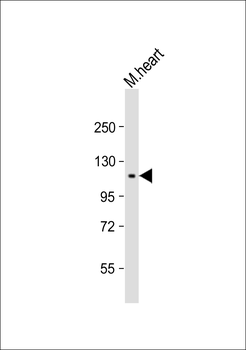
Anti-RET Antibody (Ascites) at 1:1000 dilution + mouse heart lysate. Secondary Goat Anti-mouse IgM, (H+L), Peroxidase conjugated at 1/10000 dilution. Predicted band size: 124319 Da. Blocking/Dilution buffer: 5% NFDM/TBST.
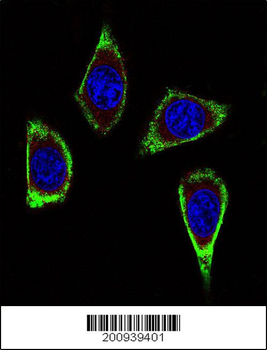
Confocal immunofluorescent analysis of RET Antibody (Ascites) with MDA-MB231 cell followed by Alexa Fluor 488-conjugated goat anti-mouse lgG (green). Actin filaments have been labeled with Alexa Fluor 555 phalloidin (red). DAPI was used to stain the cell nuclear (blue).
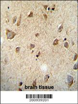
RET Monoclonal (Ascites) immunohistochemistry analysis in formalin fixed and paraffin embedded human brain tissue followed by peroxidase conjugation of the secondary antibody and DAB staining. This data demonstrates the use of the RET Monoclonal (Ascites) for immunohistochemistry. Clinical relevance has not been evaluated.
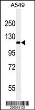
RET western blot analysis in A549 cell line lysates (35 μg/lane). This demonstrates the RET antibody detected the RET protein (arrow).
Quick Database Links
Gene Symbol
RET
UniProt
UniProt Details
− No UniProt data available
Documents Download
Datasheet
Product Information
Request a Document
Protocol Information
WB
Western Blot (IB, immunoblot)
IHC-P
Immunohistochemistry Paraffin
IF
Immunofluorescence
RET Antibody (Ascites) (orb652063)

- 0.0
Based on 0 reviews
Participating in our Biorbyt product reviews program enables you to support fellow scientists by sharing your firsthand experience with our products.
Login to Submit a ReviewAvailable Sizes
Select a size below