You have no items in your shopping cart.
Description
Research Area
Protein Biochemistry
Images & Validation
−Item 1 of 5
| Tested Applications | SDS-PAGE |
|---|---|
| Application Notes |
Key Properties
−| Source | Rabbit |
|---|---|
| Biological Origin | Rabbit |
| Isotype | IgG |
| Conjugation | Unconjugated |
| Purity | Rabbit IgG whole molecular was prepared from normal serum by a multi-step process which includes delipidation, salt fractionation and ion exchange chromatography followed by extensive dialysis against the buffer stated above. Rabbit IgG whole molecular was assayed by immunoelectrophoresis resulted in a single precipitin arc against anti-Rabbit IgG and anti-Rabbit Serum. |
Storage & Handling
−| Storage | Store vial at 4° C prior to restoration. For extended storage aliquot contents and freeze at -20° C or below. Avoid cycles of freezing and thawing. Centrifuge product if not completely clear after standing at room temperature. Rabbit IgG whole molecule is stable for several weeks at 4° C as an undiluted liquid. Dilute only prior to immediate use. |
|---|---|
| Form/Appearance | Lyophilized |
| Buffer/Preservatives | Preservative: 0.01% (w/v) Sodium Azide; Buffer: 0.02 M Potassium Phosphate, 0.15 M Sodium Chloride, pH 7.2 |
| Concentration | 10.0 mg/mL |
| Expiration Date | 6 months from date of receipt. |
| Disclaimer | For research use only |
Alternative Names
−Rabbit immunoglobulin G
Similar Products
−
Goat Rabbit IgG(H+L) Antibody, FITC conjugated [orb688925]
FC, IF, IHC
Rabbit
Goat
Polyclonal
FITC
300 μl- Item 1 of 19
Goat Anti-Rabbit IgG H&L, HRP conjugated [orb572747]
ELISA, IHC-Fr, IHC-P, WB
Rabbit
Rabbit
Goat
Polyclonal
HRP
100 μl, 1 ml - Item 1 of 23
Multi-rAb Polymer HRP-Goat Rabbit Recombinant Secondary Antibody (H+L) [orb2302681]
IHC
Rabbit
Goat
Recombinant
HRP
3 x 5 ml, 5 ml, 10 x 5 ml - Item 1 of 18
Goat Anti-Rabbit IgG H&L, Biotin conjugated [orb868233]
ELISA, FC, ICC, IF, IHC-Fr, IHC-P, WB
Rabbit
Goat
Polyclonal
Biotin
100 μl, 1 ml - Item 1 of 5
Goat Anti-Rabbit IgG (H+L) Antibody (HRP) [orb1176161]
ELISA, IHC-P, IP, WB
Rabbit
Goat
Polyclonal
HRP
20 μl, 100 μl


Quality Guarantee
Explore bioreagents carefree to elevate your research. All our products are rigorously tested for performance. If a product does not perform as described on its datasheet, our scientific support team will provide expert troubleshooting, a prompt replacement, or a refund. For full details, please see our Terms & Conditions and Buying Guide. Contact us at [email protected].
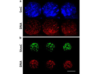
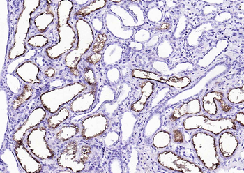
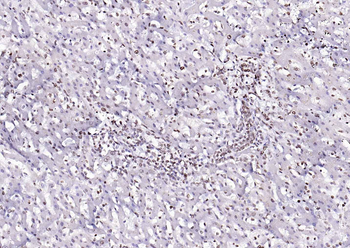
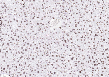
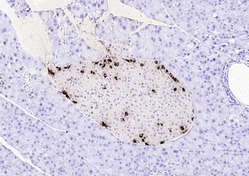
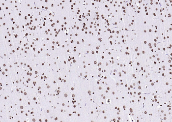
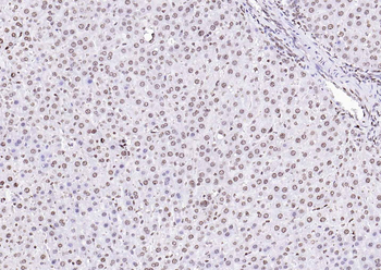
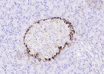
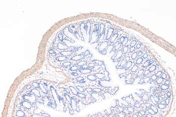
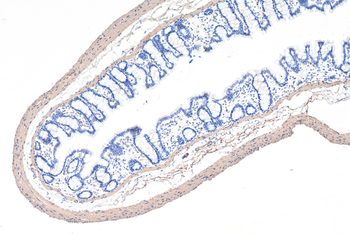
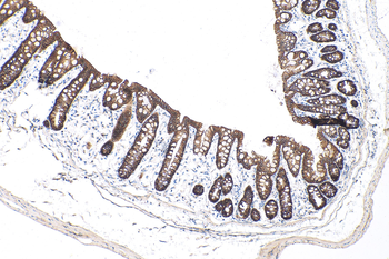
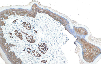
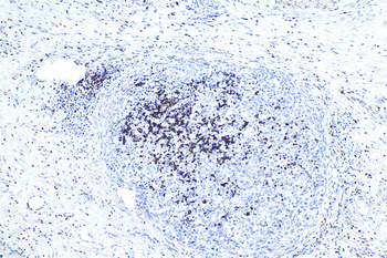
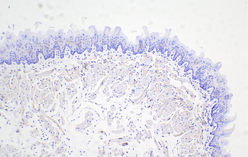
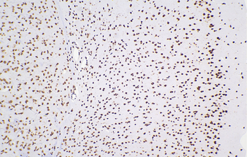
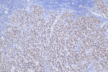
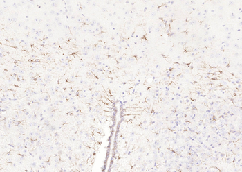
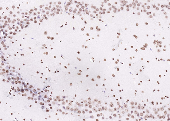
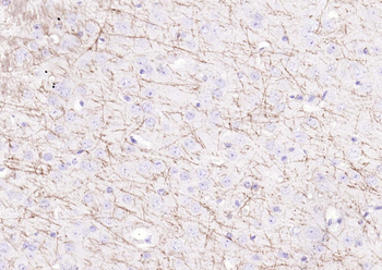
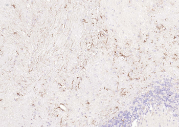
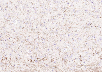
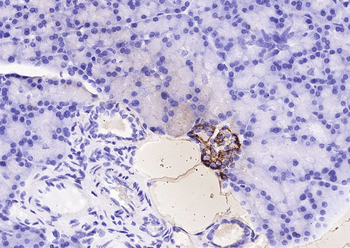
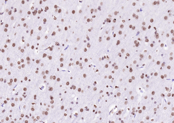
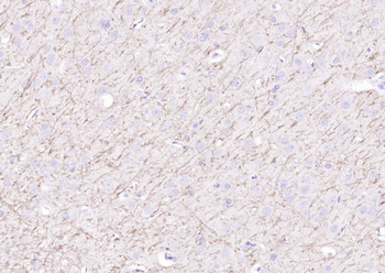
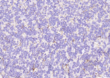
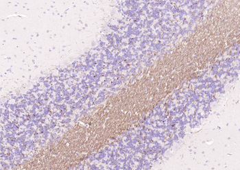
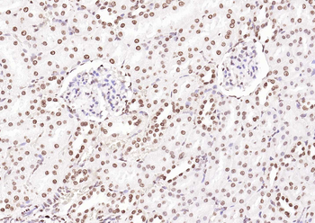
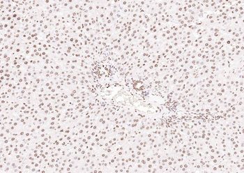
Distribution of 5-methylcytosine (5mC) and 5-hydroxymethylcytosine (5hmC) within the pronucleus. Single images were obtained at different positions of the same pronucleus from z-stacks to illustrate the distribution of 5mC and 5hmC. The pattern of distribution of 5mC and 5hmC differs largely, with 5hmC being distributed homogeneously, while 5mC was concentrated in specific DNA regions. 5mC and 5hmC were evaluated in different zygotes. Zygotes were incubated with the non-immune control antibody rabbit IgG (p/n orb2652747).
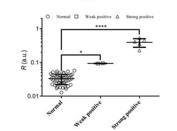
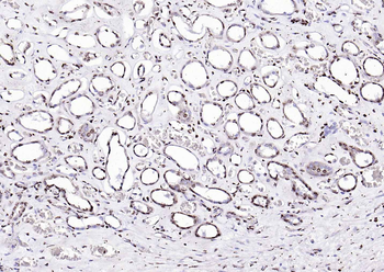
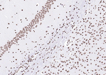
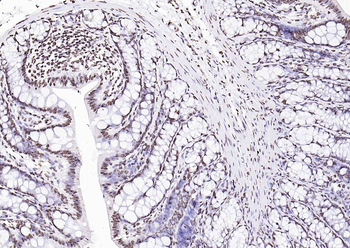
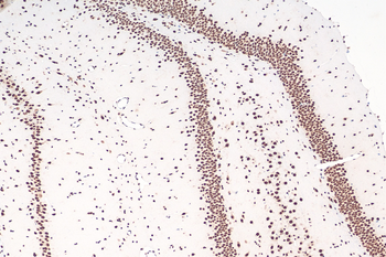
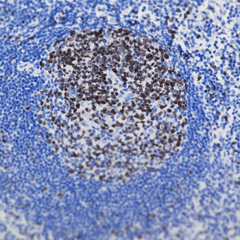
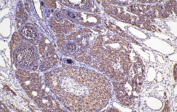
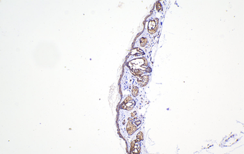
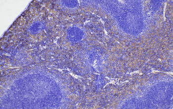
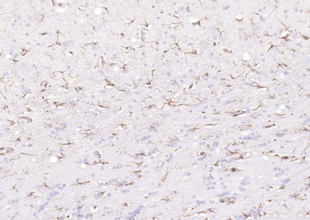
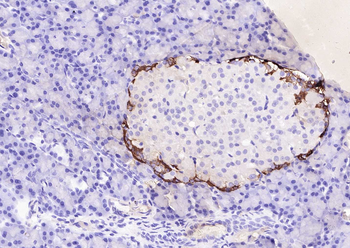
Lateral flow test results for 58 serum samples, including 51 normal and 7 positive samples. [Symbol legend: (*) P < 0.05, (****) P < 0.0001 (one-way analysis of variance and Fisher's least significant difference test).] Mouse anti-human IgG antibody and rabbit IgG [p/n orb2652747] were combined, the mixture was dispensed onto the conjugate pad and dried.
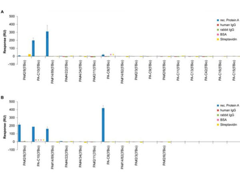
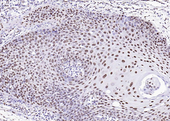
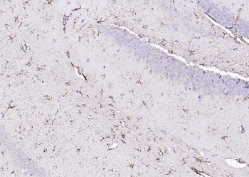
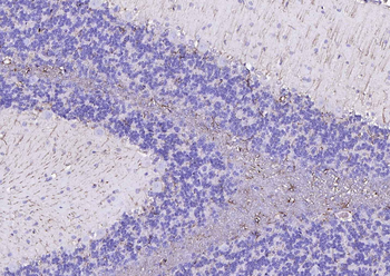
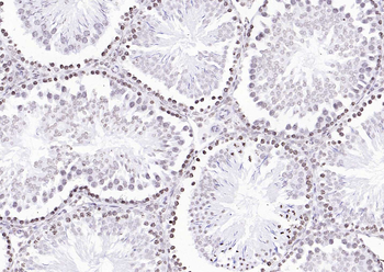
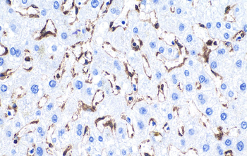
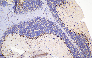
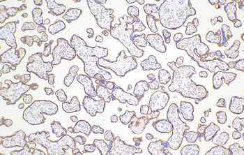
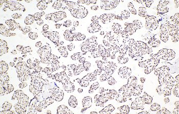
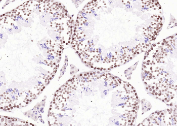
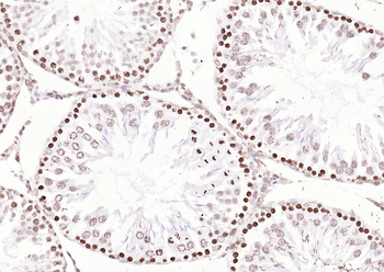
Representative sequences of the 15 identified groups from the NGS pool were screened for their individual binding abilities to Protein A. Comparative SPR-based interaction analyses were performed with the Biacore X100 instrument. Biotinylated aptamers were immobilized via the 5'-end (A) or 3'-end (B) on the streptavidin-modified sensor surface and 1000 nM Protein A was injected for binding. The sensor responses from the end of the binding phases (after 300 s) are shown. In addition, cross-specificities to other proteins were analyzed. Asterisks indicate if certain interactions have not been investigated. Rabbit IgG (p/n orb2652747).
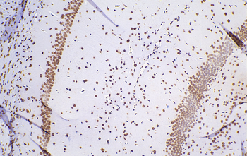
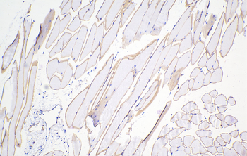
SDS-PAGE of Rabbit IgG Whole Molecule Rhodamine Conjugated (p/n orb346302). Lane M: 3 µl Opal Prestained Marker. Lane 1: Reduced Rabbit IgG Whole Molecule Rhodamine Conjugated (p/n orb346302). Lane 2: Reduced Rabbit IgG F(ab) Fragment (p/n orb2652745). Lane 3: Reduced Rabbit IgG F(c) Fragment (p/n orb346310). Lane 4: Reduced Rabbit IgM Whole Molecule (p/n orb346313). Load: 1 µg for F(ab) and F(c); 1.2 µg for IgG and IgM. Predicted/Observed size: IgG at 50 and 25 kDa; F(c) at 25 kDa; F(ab) at 25 kDa; IgM at 70 and 23 kDa. Observed F(c) Fragment migrates slightly higher.
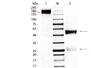
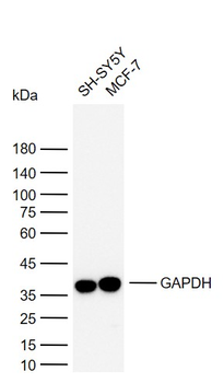
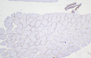
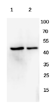
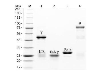
SDS-PAGE of Rabbit IgG Whole Molecule. Lane 1: Non-reduced Rabbit IgG Whole Molecule. Lane 2: 5 µl OPAL Pre-stained Marker. Lane 3: Reduced Rabbit IgG Whole Molecule. Load: 1 µg per lane. Predicted/Observed size: Non-reduced at 150-170 kDa, Reduced at 55, 25 kDa.
Documents Download
Datasheet
Product Information
Request a Document
Protocol Information
Protein Handling and Storage Guide
Protein Handling Guide
SDS-PAGE
Sodium Dodecyl Sulphate PolyAcrylamide Gel Electrophoresis
Rabbit IgG (orb2652747)

- 0.0
Based on 0 reviews
Participating in our Biorbyt product reviews program enables you to support fellow scientists by sharing your firsthand experience with our products.
Login to Submit a Review