You have no items in your shopping cart.
Rabbit SND1 Antibody
SKU: orb1525829
Description
Research Area
Cancer Biology, Immunology & Inflammation
Images & Validation
−Item 1 of 2
| Tested Applications | IHC, IP, WB |
|---|---|
| Dilution Range | WB - 1:2,000 - 1:10,000; IP - 2 - 5 µg/mg lysate; IHC - 1:100 - 1:500. Epitope retrieval with citrate buffer pH 6.0 is recommended for FFPE tissue sections. |
| Reactivity | Human, Mouse |
| Predicted Reactivity | Rat |
| Application Notes |
Key Properties
−| Antibody Type | Primary Antibody |
|---|---|
| Host | Rabbit |
| Clonality | Polyclonal |
| Isotype | IgG |
| Immunogen | Between 125 and 175 |
| Target | SND1 |
| Purification | Antigen Affinity Purified |
| Conjugation | Unconjugated |
Storage & Handling
−| Storage | 2 - 8°C |
|---|---|
| Form/Appearance | Liquid |
| Buffer/Preservatives | Tris-buffered Saline containing 0.1% rAlbumin and 0.09% Sodium Azide |
| Concentration | 200 µg/ml |
| Expiration Date | 12 months from date of receipt. |
| Disclaimer | For research use only |
Alternative Names
−100 kDa coactivator; EBNA2 coactivator p100; p100; p100 co-activator; staphylococcal nuclease domain-containing protein 1; TDRD11; testis tissue sperm-binding protein Li 82P; tudor domain-containing protein 11; Tudor-SN
Similar Products
−- Item 1 of 5
SND1 Rabbit Polyclonal Antibody [orb745969]
ELISA, FC, ICC, IF, IHC, WB
Human, Mouse, Rat
Rabbit
Polyclonal
Unconjugated
100 μg - Item 1 of 4
TudorSN rabbit pAb Antibody [orb766689]
ELISA, IF, IHC, WB
Human, Mouse, Rat
Polyclonal
Unconjugated
100 μl - Item 1 of 2
SND1 Rabbit Polyclonal Antibody [orb574269]
IHC, WB
Bovine, Canine, Equine, Guinea pig, Mouse, Rabbit, Rat, Zebrafish
Human
Rabbit
Polyclonal
Unconjugated
100 μl - Item 1 of 2
SND1 Rabbit Polyclonal Antibody [orb631364]
ELISA, IF, IHC, WB
Human, Mouse, Rat
Rabbit
Polyclonal
Unconjugated
50 μg, 100 μg - Item 1 of 1


Quality Guarantee
Explore bioreagents carefree to elevate your research. All our products are rigorously tested for performance. If a product does not perform as described on its datasheet, our scientific support team will provide expert troubleshooting, a prompt replacement, or a refund. For full details, please see our Terms & Conditions and Buying Guide. Contact us at [email protected].

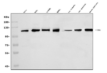
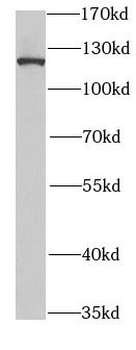
Detection of human and mouse SND1 by western blot (h&m) and immunoprecipitation (h). Samples: Whole cell lysate from HeLa (5, 15 and 50 µg for WB; 1 mg for IP, 20% of IP loaded) , HEK293T (T; 50 µg) , and mouse NIH 3T3 (M; 50 µg) cells. Antibodies: Affinity purified rabbit anti-SND1 antibody orb1525829 used for WB at 0.04 µg/ml (A) and 0.4 µg/ml (B) and used for IP at 3 µg/mg lysate. SND1 was also immunoprecipitated by rabbit anti-SND1 antibody, which recognizes a downstream epitope. Detection: Chemiluminescence with exposure times of 30 seconds (A) and 10 seconds (B).
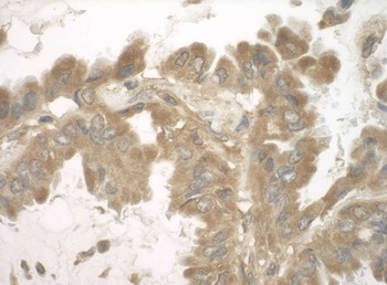
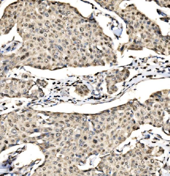
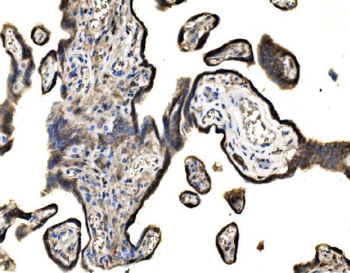
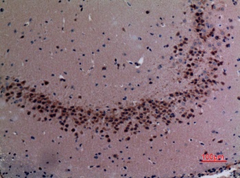
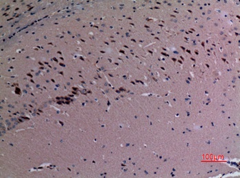
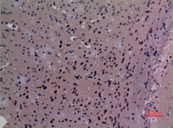
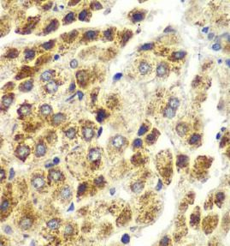
Detection of human SND1 by immunohistochemistry.Sample: FFPE section of human lung carcinoma. Antibody: Affinity purified rabbit anti-SND1 (Cat. No. orb1525829) used at a dilution of 1: 200 (1µg/ml). Detection: DAB
Quick Database Links
UniProt Details
− No UniProt data available
NCBI Reference Sequences
−Associated Accession Numbers
Curated reference sequences for the gene transcript and protein product| Protein | NP_055205.2 |
|---|
Documents Download
Datasheet
Product Information
Request a Document
Protocol Information
WB
Western Blot (IB, immunoblot)
IHC
Immunohistochemistry
IP
Immunoprecipitation
Rabbit SND1 Antibody (orb1525829)

- 0.0
Based on 0 reviews
Participating in our Biorbyt product reviews program enables you to support fellow scientists by sharing your firsthand experience with our products.
Login to Submit a ReviewAvailable Sizes
Select a size below