You have no items in your shopping cart.
PDIA3 Antibody
SKU: orb1264508
Featured
Description
Research Area
Signal Transduction
Images & Validation
−Item 1 of 4
| Tested Applications | FC, IHC-P, WB |
|---|---|
| Reactivity | Human |
| Predicted Reactivity | Bovine |
| Application Notes |
Key Properties
−| Antibody Type | Primary Antibody |
|---|---|
| Host | Rabbit |
| Clonality | Polyclonal |
| Isotype | Rabbit Ig |
| Immunogen | This PDIA3 antibody is generated from rabbits immunized with a KLH conjugated synthetic peptide between 192-220 amino acids from the Central region of human PDIA3. |
| Target | PDIA3 |
| Molecular Weight | 57 kDa |
| Purification | This antibody is prepared by Saturated Ammonium Sulfate (SAS) precipitation followed by dialysis |
| Conjugation | Unconjugated |
Storage & Handling
−| Storage | Maintain refrigerated at 2-8°C for up to 2 weeks. For long term storage store at -20°C in small aliquots to prevent freeze-thaw cycles. |
|---|---|
| Form/Appearance | Liquid |
| Buffer/Preservatives | Supplied in PBS with 0.09% (W/V) sodium azide. |
| Concentration | batch dependent |
| Expiration Date | 12 months from date of receipt. |
| Disclaimer | For research use only |
Alternative Names
−Protein disulfide-isomerase A3, 58 kDa glucose-regulated protein, 58 kDa microsomal protein, p58, Disulfide isomerase ER-60, Endoplasmic reticulum resident protein 57, ER protein 57, ERp57, Endoplasmic reticulum resident protein 60, ER protein 60, ERp60, PDIA3, ERP57, ERP60, GRP58
Similar Products
−- Item 1 of 8
PDIA3 Rabbit Polyclonal Antibody [orb585694]
IHC, IP, WB
Bovine, Canine, Equine, Goat, Guinea pig, Mouse, Rabbit, Rat, Sheep, Zebrafish
Human
Rabbit
Polyclonal
Unconjugated
100 μl - Item 1 of 4
PDIA3 Antibody (Center) [orb1931335]
FC, IHC-P, WB
Bovine
Human
Rabbit
Polyclonal
Unconjugated
100 μl, 50 μl - Item 1 of 4
PDIA3 Antibody (C-term) [orb1931336]
FC, IHC-P, WB
Human, Mouse, Rat
Rabbit
Polyclonal
Unconjugated
100 μl, 50 μl - Item 1 of 4
ERp57 rabbit pAb Antibody [orb766825]
ELISA, IF, IHC, WB
Human, Mouse, Rat
Polyclonal
Unconjugated
100 μl - Item 1 of 5
ERp57/PDIA3 Rabbit Polyclonal Antibody [orb334504]
FC, ICC, IF, IHC, IHC-Fr, WB
Human, Mouse, Rat
Rabbit
Polyclonal
Unconjugated
100 μg

Quality Guarantee
Explore bioreagents carefree to elevate your research. All our products are rigorously tested for performance. If a product does not perform as described on its datasheet, our scientific support team will provide expert troubleshooting, a prompt replacement, or a refund. For full details, please see our Terms & Conditions and Buying Guide. Contact us at [email protected].
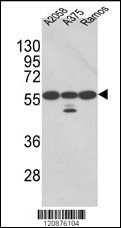
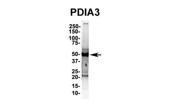
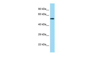
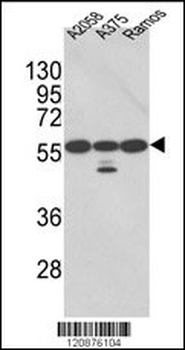
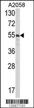
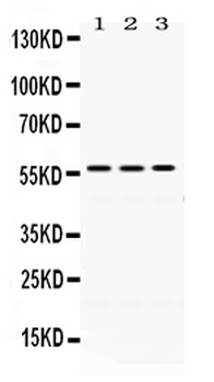
Western blot analysis of PDIA3 Antibody in A2058, A375, Ramos cell line lysates (35 ug/lane) (2ug/ml)
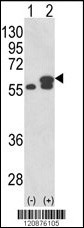
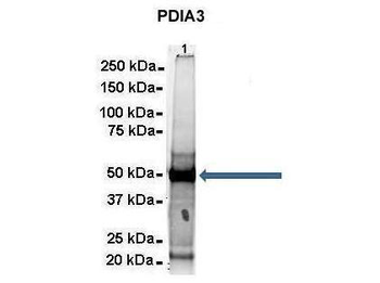
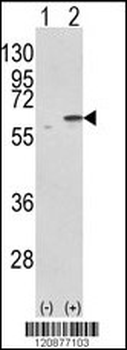
Western blot analysis of PDIA3 using rabbit polyclonal PDIA3 Antibody using 293 cell lysates (2 ug/lane) either nontransfected (Lane 1) or transiently transfected with the PDIA3 gene (Lane 2).
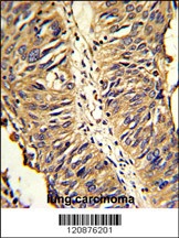
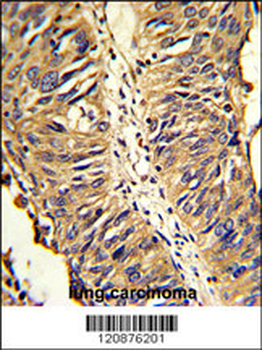
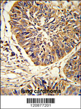
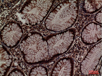
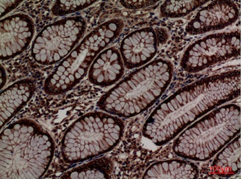
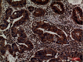
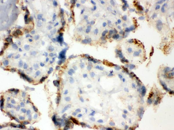
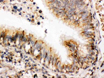
Formalin-fixed and paraffin-embedded human lung carcinoma reacted with PDIA3 Antibody, which was peroxidase-conjugated to the secondary antibody, followed by DAB staining.
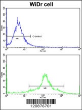
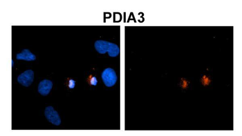
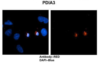
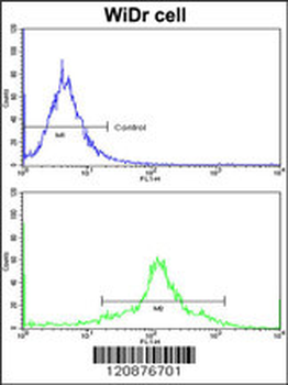
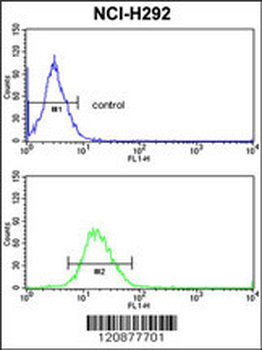
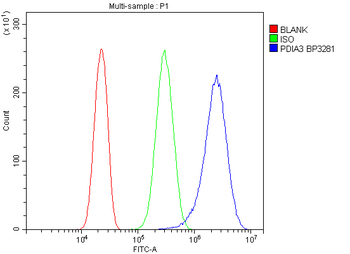
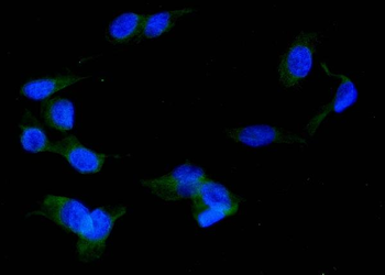
Flow cytometric analysis of widr cells using PDIA3 Antibody (bottom histogram) compared to a negative control cell (top histogram) FITC-conjugated goat-anti-rabbit secondary antibodies were used for the analysis.
Documents Download
Datasheet
Product Information
Request a Document
Protocol Information
WB
Western Blot (IB, immunoblot)
IHC-P
Immunohistochemistry Paraffin
FC
Flow Cytometry
PDIA3 Antibody (orb1264508)

- 0.0
Based on 0 reviews
Participating in our Biorbyt product reviews program enables you to support fellow scientists by sharing your firsthand experience with our products.
Login to Submit a ReviewAvailable Sizes
Select a size below