You have no items in your shopping cart.
NpFR1
SKU: orb1714148
Description
Images & Validation
−Item 1 of 5
Key Properties
−| MW | 431.45 |
|---|---|
| Formula | C23H21N5O4 |
| SMILES | C1(N(C(C2=CC5=C(C3=CC=CC1=C23)N(C4=NC(NC(C4=N5)=O)=O)CCC)=O)CCCC)=O |
| Solubility | Soluble in DMSO |
Storage & Handling
−| Storage | -20°C |
|---|---|
| Expiration Date | 12 months from date of receipt. |
| Hazard Information | Classification: Caution: Substance not yet fully tested. Safety Phrases: S22 - Do not breathe dust S24/25 - Avoid contact with skin and eyes S36/37/39 - Wear suitable protective clothing, gloves and eye/face protection |
| Disclaimer | For research use only |
Similar Products
−Quality Guarantee
Explore bioreagents carefree to elevate your research. All our products are rigorously tested for performance. If a product does not perform as described on its datasheet, our scientific support team will provide expert troubleshooting, a prompt replacement, or a refund. For full details, please see our Terms & Conditions and Buying Guide. Contact us at [email protected].
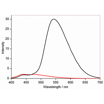
Fluorescence behavior of NpFR1 in the oxidized (black) and reduced (red) forms, using 50 μM. Excitation: 405 or 488 nm (not shown). Emission: 490 – 600 nm, with peak at 545 nm.
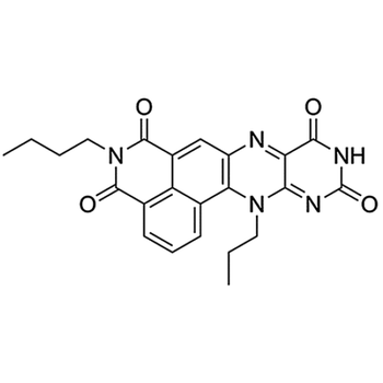
Chemical structure of NpFR1, a reversible fluorescence intensity-based redox sensor.
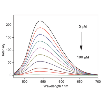
Fluorescence emission of NpFR1 (5 mM, lex = 405 nm) with the incremental addition of sodium dithionite. All spectra were acquired in HEPES buffer (100 mM, pH 7.4).
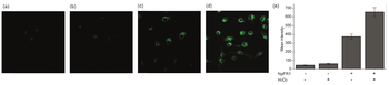
Imaging of NpFR1 in 3T3-L1 preadipocytes treated with (a) vehicle control (b) H2O2 (100 mM, 2 min), (c) NpFR1 (50 mM, 2 h) and (d) NpFR1 (50 mM, 2 h) followed by H2O2 (100 mM, 2 min). Scale bar represents 50 mm, lex = 405 nm. (e) Integrated emission from 510 nm to 610 nm. Values are the mean ratio generated from the intensity from five fields of cells. Error bars represent standard error measurement (s.e.m.).
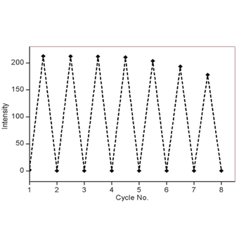
Fluorescence response of NpFR1 to cycles of oxidation and reduction. Reduction was achieved with sodium dithionite (100mM) followed by re-oxidation with 250mM H2O2. Spectra were recorded 5 min after the addition of reducing and oxidising agents. All spectra were acquired in HEPES buffer (100 mM, pH 7.4).
Compound Identifiers
PubChem CID
Key Properties
− No computed properties available.
Documents Download
Datasheet
Product Information
Request a Document
Protocol Information
NpFR1 (orb1714148)

- 0.0
Based on 0 reviews
Participating in our Biorbyt product reviews program enables you to support fellow scientists by sharing your firsthand experience with our products.
Login to Submit a ReviewAvailable Sizes
Select a size below